
Figure 5.
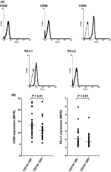
Expression of costimulatory molecules on CD14+ HLA‐DR − cells. Flow cytometry was performed as described in the Materials and Methods. Representative data of CD14+ HLA‐DR − cells obtained from one cancer patient (A). Expression of CD86 and PD‐L1 on CD14+ HLA‐DR − and CD14+ HLA‐DR + cells in peripheral blood mononuclear cells (PBMC) obtained from patients with squamous cell carcinoma of the head and neck (B). Lines indicate mean values. MFIR, mean fluorescence intensity ratio.