
Figure 9.
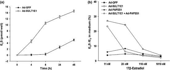
Estrogen sulfation by Ad‐SULT1E1 and/or Ad‐PAPSS1. The formation of sulfated estrogen was analyzed by alkaline–chloroform extraction. MCF‐7 cells were cultured in a 24‐well plate and infected with Ad‐GFP, Ad‐SULT1E1, Ad‐PAPSS1 or Ad‐SULT1E1 mixed with Ad‐PAPSS1 for 48 h. Then the cells were incubated with 10 nM [3 H] E 2 in the absence or presence of 1, 10, 100, 1000 nM cold estrogen for the indicated time. Sulfated estrogen in the medium was measured by liquid scintillation counting as described in Materials and Methods. The E 2 S formation in time‐course is shown in (a). The ratio of E 2 S to E 2 in cultured medium is shown in (b).