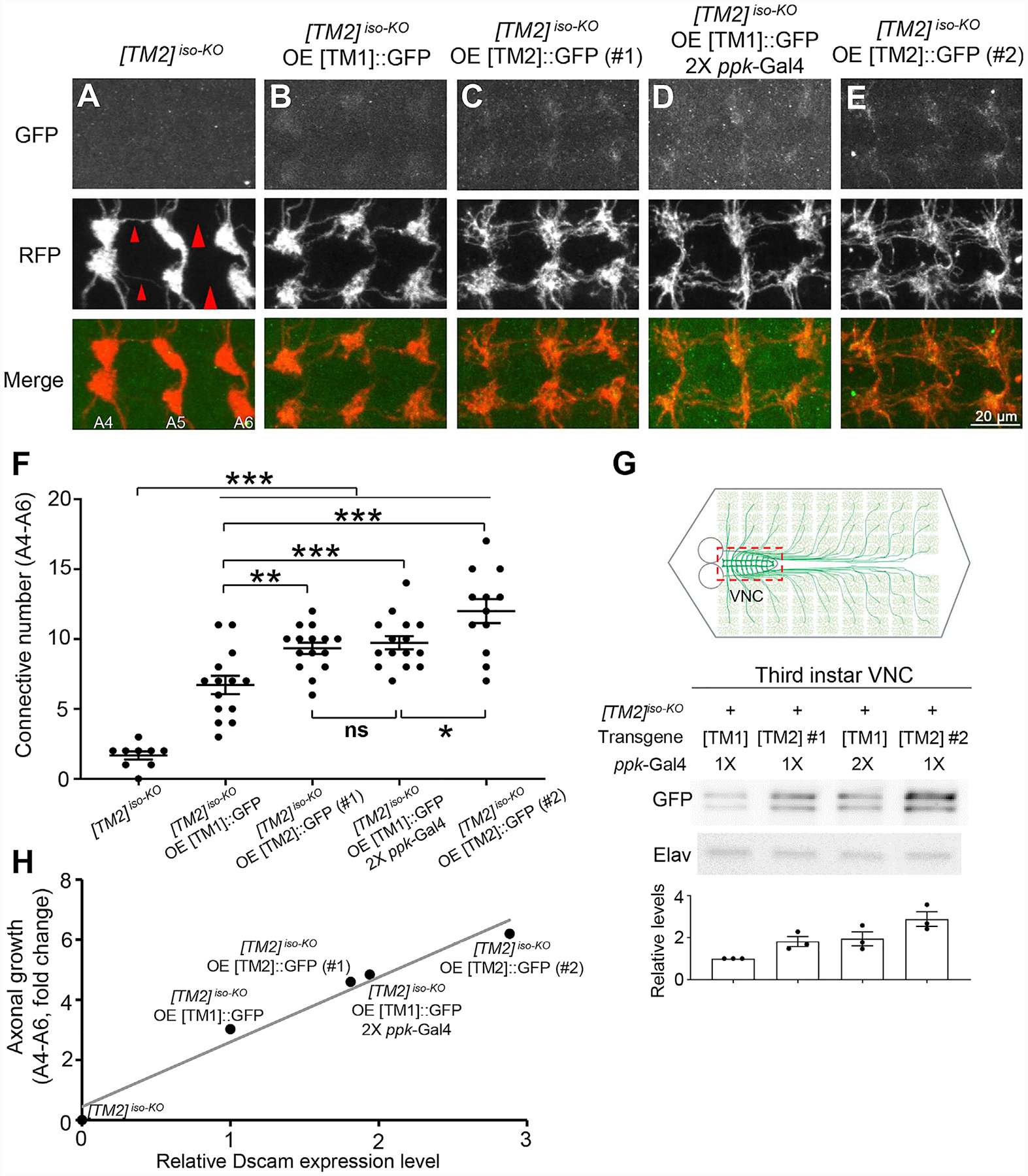
Figure 5. Dendrite-Specific Localization Restrains Endogenous Dscam[TM1] from Functioning in Axons.

(A) Dscam[TM2]iso-KO dramatically impairs the axon terminal growth in C4 da neurons. The large red arrowheads point to the sites where longitudinal axon tracts are broken, and the small arrowheads point to where the tracts are thinned.
(B–E) Overexpression of a Dscam[TM1], Dscam[TM2]#1, or Dscam[TM2]#2 transgenes with the ppk-Gal4 driver leads to axon localization of Dscam isoforms at different levels and mitigates the axonal defect in Dscam[TM2]iso-KO to different extents.
(F) Quantification of the number of C4 da axon connectives in segments A4-A6.
(G) Quantification of transgenic Dscam::GFP levels in C4 da axon terminals. The experiments were done in Dscam[TM2]iso-KO larvae. As shown in the schematic, UAS-Dscam::GFP transgenes were expressed in C4 da neurons with the ppk-Gal4 driver. VNCs (indicated by the dashed red box in the top panel), which contained transgenic Dscam::GFP in C4 da axon terminals, were dissected out from third-instar larvae for western blotting.
(H) The rescue of the axonal defect caused by the loss of Dscam is proportional to the level of transgenic Dscam in C4 da axon terminals, regardless of the isoform. The relative Dscam expression levels were determined by western blotting of CNS lysates from larvae overexpressing [TM1]::GFP or [TM2]::GFP by ppk-Gal4 and plotted against the rescue effect of the transgene. Linear regression, R2 = 0.969.
