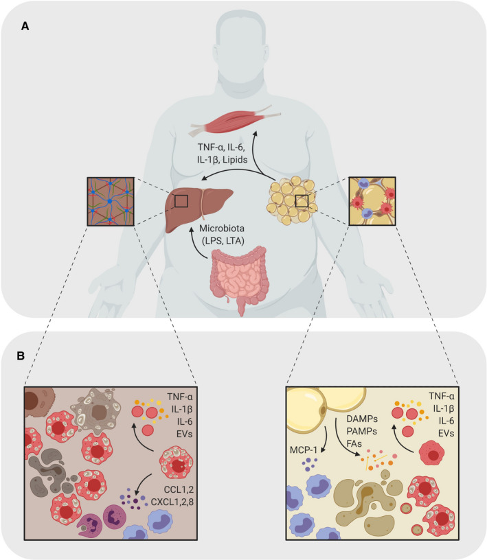
Figure 1.

Role of macrophages in obesity‐driven NAFLD. A, Inflammatory macrophage activation (shown in red) promoting the progression of NAFLD involves a cross‐talk of AT, liver, gut and skeletal muscles. The inflammatory state in the AT leads to secretion of lipids and pro‐inflammatory cytokines into the blood stream where they travel to other metabolic organs, such as the skeletal muscles and the liver. Here, they induce an inflammatory state that can lead to metabolic diseases caused by chronic low‐grade inflammation. In obesity, the liver also receives LPS and lipoteichoic acid (LTA) from the gut, which also contribute to inflammation and tissue damage. B, During obesity, adipocytes stimulate monocyte infiltration by secreting MCP‐1. Adipocyte secretion of various DAMPs, PAMPs and FAs mediate a pro‐inflammatory polarization and differentiation of ATMs and monocytes. Within the liver, excess lipids, apoptitic bodies and AT‐derived cytokines induce an inflammatory environment with pro‐inflammatory hepatic macrophages and foam cells (shown in red). Their respond to the constant stimuli maintains the inflammatory state of the tissue by secreting pro‐inflammatory factors and neutrophil‐ and monocyte (shown in purple) attractants.
