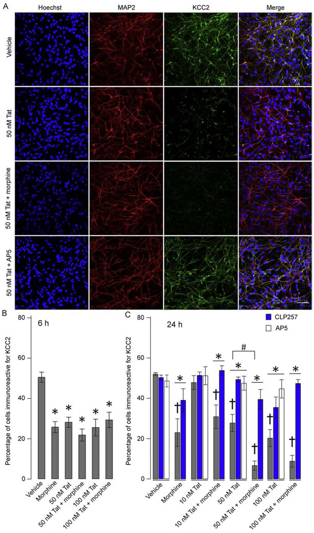
Figure 4. hNeurons lose KCC2 immunoreactivity after exposure to HIV-Tat ± morphine.
(A) Representative images of hNeuron cultures immunolabeled for MAP2 and KCC2. Loss of KCC2 immunoreactivity can be noted in cells treated with both 50 nM Tat1-86 and 50 nM Tat1-86 + morphine. Rescue is seen in 50 nM Tat1-86 + AP5 (scale bar = 50 μm). (B, C) The percentage of cells immunoreactive for KCC2. Both 6 h (B) and 24 h (C) exposure to 50 - 100 nM Tat1-86 ± morphine results in significant decrease in KCC2 immunoreactivity compared to their respective controls (†p < 0.01; n = 6 – 7). Further, a significant interaction between morphine and Tat1-86 was found at the 50 nM Tat1-86 level after 24 h exposure (#p < 0.05). Application of CLP257 (blue) and AP5 (white) showed significant restoration of KCC2 immunoreactivity across all groups and Tat1-86 exposed groups, respectively (*p < 0.01).

