
Figure 2.
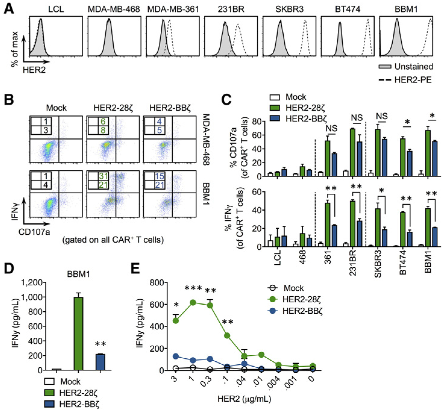
HER2-BBζ CAR T cells show reduced but antigen-specific cytokine production compared with HER2–28ζ CAR T cells. A, Histograms of HER2 expression in human cancer cell lines. LCL is an EBV-transformed human lymphoblastoid cell line. MDA-MB-468, MDA-MB-361, MDA-MB-231BR (231BR), SKBR3, and BT474 are breast cancer cell lines with varying HER2 expression. BBM1 is a patient-derived tumor line generated from the brain metastasis of a breast cancer patient. B, Representative zebra plots showing cell surface CD107a and intracellular IFNγ expression by Mock, HER2–28ζ, or HER2-BBζ CAR T cells following a 5-hour coculture with indicated tumor targets at an E:T ratio of 1:1. C, Quantification of CD107a degranulation and IFNγ production by HER2-CAR T cells using data generated from histograms as depicted in B. D, IFNγ production quantified by ELISA in supernatants from HER2-CAR T cells cultured overnight with BBM1 tumor cells at a 1:1 E:T ratio. E, IFNγ production quantified by ELISA in supernatants from Mock and HER2-CAR T cells cultured on plate-bound recombinant human HER2 at the indicated protein concentrations. *, P < 0.05; **, P < 0.01; ***, P < 0.001.