Figure 1.
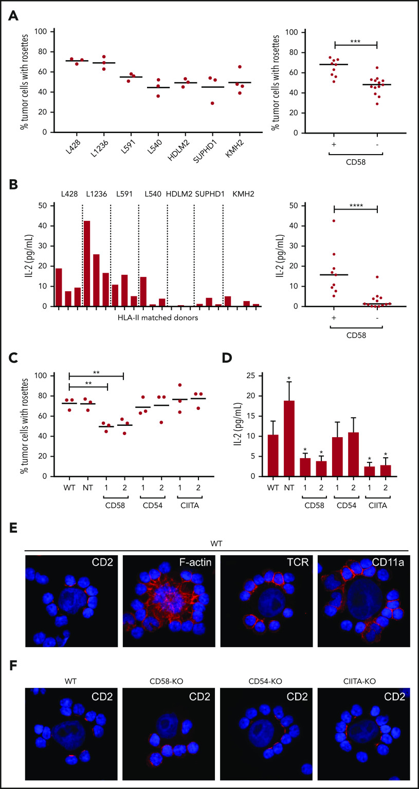
Involvement of HLA-II, CD58, and CD54 in adhesion and activation of rosetting T cells using PMBCs from at least 3 individual HLA-II–matched donors. (A) The percentage of tumor cells with rosettes was quantified after a 30-minute coculture of HL cell lines and matched PBMCs. CD58+ cell lines were L591, L1236, and L428, and the CD58− cell lines were KMH2, SUPHD1, HDLM2, and L540. (B) TCR-mediated T-cell activation was determined by measuring IL-2 (picograms per milliliter) in the culture supernatant after 18 hours of coculture with different HL cell lines. (C) The percentage of tumor cells with rosettes was quantified after a 30-minute coculture of L428 WT, nontargeting (NT) control, and 2 independently generated CD58-KO, CD54-KO, or CIITA-KO cell lines with matched PBMCs. CIITA was targeted to generate HLA-II low cells. In panels A-C, each dot or bar represents 1 independent HLA-II–matched donor used for cocultures. (D) IL-2 production of matched PBMCs in response to a coculture with WT, NT, CD58-KO, CD54-KO, or CIITA KO L428 cell lines. Data are expressed as the mean of 3 different HLA-II–matched PBMC donors ± standard deviation. (E) CD2, TCR, CD11a, and F-actin staining (red signal) of L428 cells cocultured with matched PBMCs. Nuclei were counterstained with 4′,6-diamidino-2-phenylindole (DAPI; blue signal). (F) CD2 staining of WT, CD58-KO, CD54-KO, and CIITA-KO L428 cell lines cocultured with matched PBMCs. Staining was visualized using a Leica DM4000B microscope equipped with a Leica DFC345FX Camera and LAS V4.8 software. Relocalization of CD2 can be observed as discrete red signals at the interface between T cells and tumor cells instead of the homogenous circumferential staining of entire T cells. Relocalization of F-actin in tumor cells presented as spider-web–like structures stretching out toward the T cells. Representative images are shown; original magnification ×400. Statistical significance was calculated with a 1-tailed Mann-Whitney U test if different cell lines were compared (A-B) or a 1-tailed paired Student t test if a KO was compared with the WT (C-D). *P < .05; **P < .01; ***P < .001; ****P < .0001.

