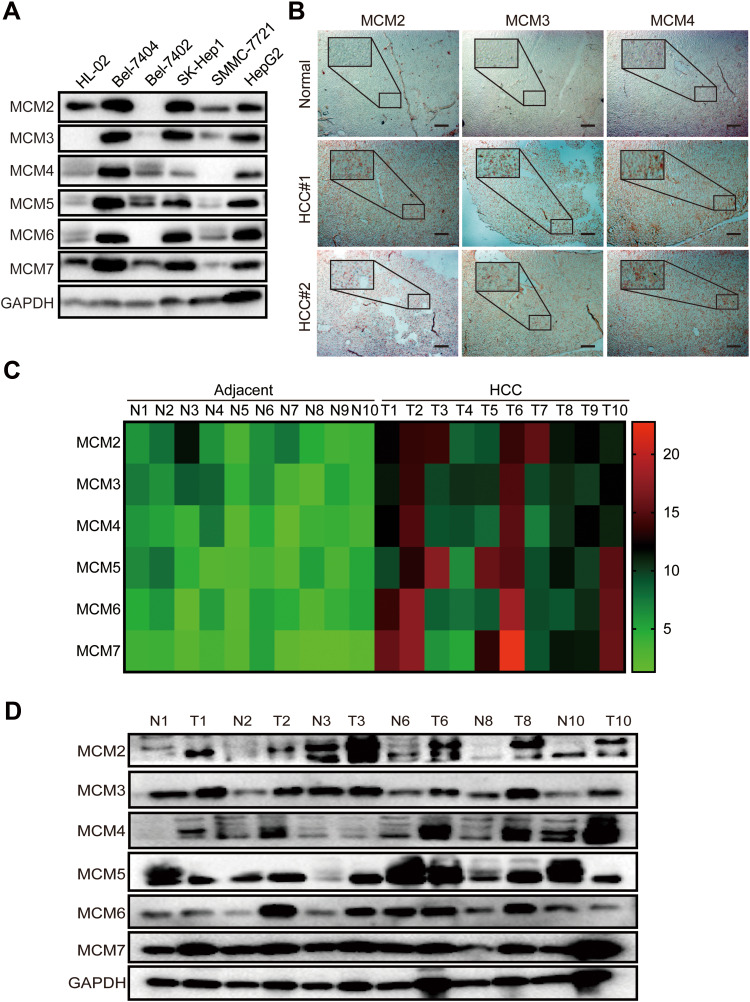
Figure 3.
MCM proteins were up-regulated in HCC cell lines and tissues. (A) Western blots assay showed the expression levels of MCM2, MCM3, MCM4, MCM5, MCM6, and MCM7 in HL-02, Bel-7404, SK-Hep1, Bel-7402, SMMC-7721, and HepG2 cell lines. (B) Representative IHC images of MCM2, MCM3, and MCM4 staining from normal liver and HCC tissues, scar bar 100μm. (C) The heat-map exhibits the differentially expressed MCM between 10 pairs of HCC tissues and adjacent normal tissues measured by RT-qPCR assay. (D) Western blots of the expression levels of MCM proteins between 6 pairs of HCC tissues and adjacent normal tissues.

