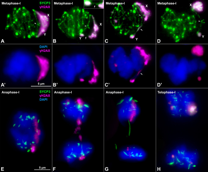
Fig 5. Sex chromosome segregation.
Squashed spermatocytes labelled with antibodies against SYCP3 (green) and γH2AX (magenta) and counterstained with DAPI (blue). Non-homologous segments of the sex chromosomes (X, Y) are indicated. A-D’. Metaphase-I. Different configurations of the sex bivalent can be observed. The neo-PAR always shows a chiasma (arrowheads), however, the non-homologous segments can be associated with each other by forming a common chromatin mass (A-A’), or through a SYCP3-positive filament (arrow) (see enlarged detail on the top) (B-B’) or a γH2AX-positive filament (arrow) (C-C’). Alternatively, these segments can appear completely separated (D-D’). E-G. Anaphase-I. γH2AX connections (E) and/or SYCP3 (F, G) filaments are observed between segregating chromosomes during early anaphase, resembling the associations observed at metaphase-I. At telophase-I (H), no lagging or mis-segregated chromosomes are observed.

