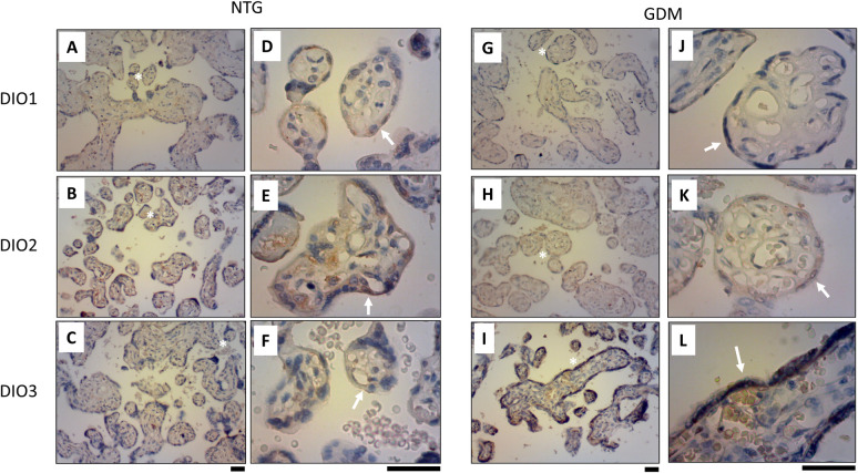
Fig 3. GDM effect on DIO2 and DIO 3 expression in placental tissue.
DIO2 and DIO3 immunodetection were realized by immunohistochemistry (IHC) in normal glucose tolerant (NTG, A-F) and gestational diabetes mellitus (GDM, G-L) pregnancies. In A-C and G-I 400x magnification, and in D-F and J-L 1000x magnification. White asterisk (*) in A-C and G-I indicates a magnification point in D-F and J-L, respectively. White arrow in D-F and J-L indicates syncytiotrophoblast in the placental tissue. Black bar: 50μm. N = 5 per group.

