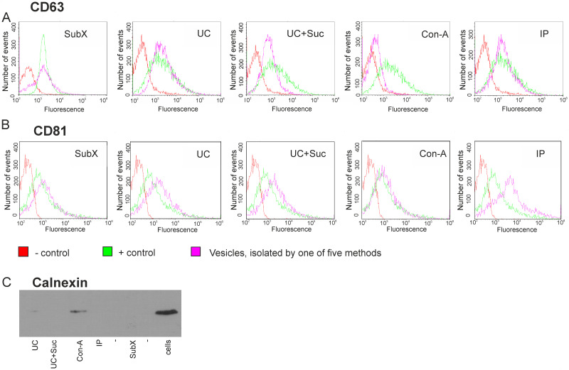
Fig 7. Analysis for positive and negative exosome markers in the samples of vesicles isolated by 5 methods.
Flow cytometry analysis of isolated vesicles for the surface expression of CD63 (A) and CD81 (B) tetraspanins classically used as exosome markers. Immunobeads blocked with BSA and stained with anti-CD63 or CD81 antibodies were used as negative control (–control). The exosomal standard included in the exosome cytometric assay kit (Lonza) was used as a positive control (+ control). Western blot analysis for exosome negative marker, calnexin, in isolated samples of vesicles and U-87 MG cell lysate sample (C). Designation of the isolation methods: SubX™ technology; Ultracentrifugation–UC; ultracentrifugation in the cushion of 30% sucrose—UC + Suc, precipitation of lectin aggregates—Con-A; immunoprecipitation–IP.

