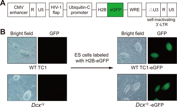
Figure 3. Lentiviral transduction of ESCs.
(A) Diagram of the lentiviral vector FUGW-H2B-eGFP (adapted from25) used to fluorescently label ESCs. Only the relevant portions of the plasmid are shown. (B) Representative bright field and fluorescence microscopy images of wild-type TC1 ES cells and Dcx-/y ES cells before and after lentiviral integration of H2B–eGFP (original magnification, 100×). The ES cells shown stably express H2B–eGFP and were repeatedly imaged during the study with similar results. Also note the ESC morphology. The ESC clones shown are approximately 100–150 μm long. Images were acquired with a Nikon Diaphot 300 inverted fluorescence phase contrast microscope.

