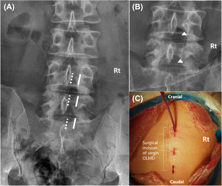
Fig. 1.
Location of the surgical portal. a The viewing and working surgical ports (bold line) were made right inside the medial pedicular line of the target segment and above the intact upper and lower laminas. These were located more lateral than the ports made during conventional biportal endoscopic discectomy (dotted line). b Direct access to the lamina and the facet joint was made to complete the red vision discectomy with minimal laminotomy (triangle). c Clinical photographs show independent surgical ports on the outside of the previous midline incision site

