. 2020 Nov 24;9:e60729. doi: 10.7554/eLife.60729

Figure 2. Segmentations of the amygdalae and connected regions defined by the top 20% streamline counts.

Figure 1a shows the lateral view of the sagittal plane and 1b the medial view. The same eight regions had the highest streamline counts to the amygdalae bilaterally.

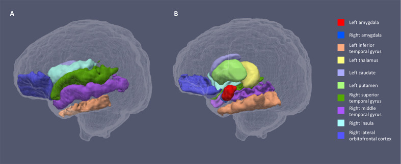
Figure 2.

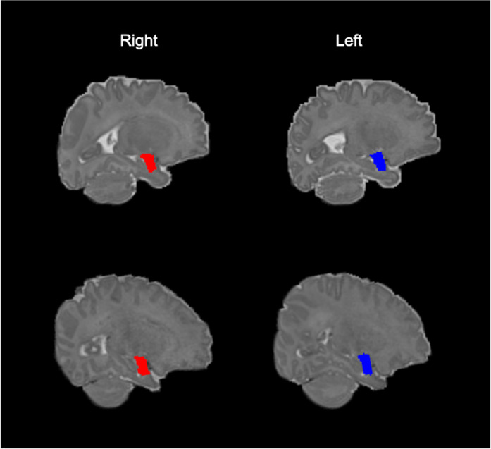
Figure 2—figure supplement 1. Example segmentations of the amygdala in native space.

Figure 2—figure supplement 1.

Right and left amygdala segmentations from a preterm infant at term equivalent age (upper row) and an infant born at term (lower row), viewed in native space.