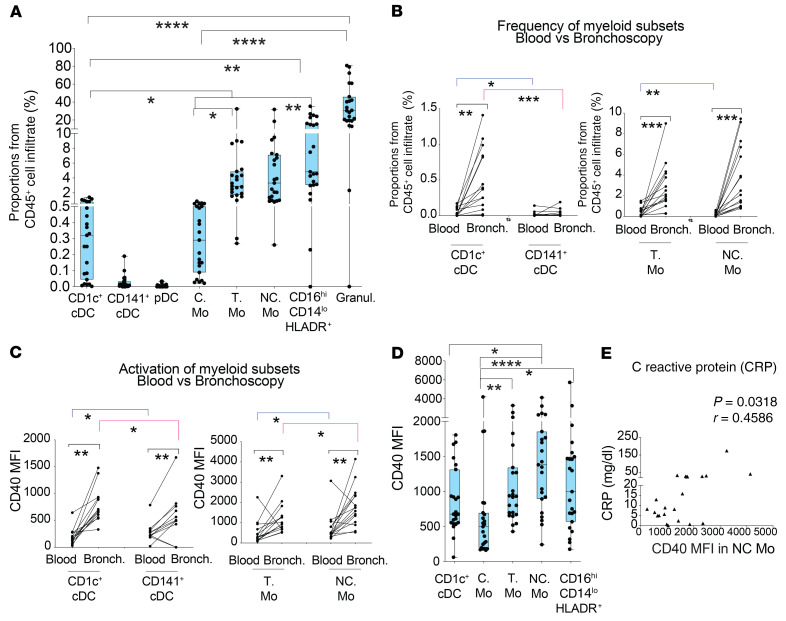
Figure 5. Characterization of myeloid cell subsets present in bronchoscopy infiltrates from COVID-19 patients with ARDS.
(A) Box-and-whiskers plots showing percentages of the indicated cell populations in the hematopoietic CD45+ infiltrate present in bronchoscopy mucus samples from severe COVID-19 patients (n = 23) presenting ARDS and receiving IMV at ICU. Error bars represent maximum and minim values. Statistical differences between proportions of cell populations within the same infiltrates were calculated using Friedman’s test for multiple comparisons. *P < 0.05; **P < 0.01; ****P < 0.0001. (B and C) Frequencies (B) and CD40 MFI (C) of CD1c+ and CD141+ cDCs (left) and T and NC Mo (right) in paired blood and bronchoscopy samples from COVID-19 patients presenting with ARDS (n = 15). Statistical significance of differences in frequencies between paired blood vs. bronchoscopy samples (black) or between different cell subsets within either blood (blue) or bronchoscopy infiltrates (pink) was calculated using a 2-tailed matched pairs Wilcoxon’s test. *P < 0.05; **P < 0.01; ***P < 0.001. (D) Box-and-whiskers plots representing comparison of CD40 MFI on the indicated myeloid cell populations present in the bronchoscopy infiltrates of total critical G3 COVID-19 patients. Error bars represent maximum and minimum values. Statistical significance of differences was calculated using Friedman’s test for multiple comparisons. *P < 0.05; **P < 0.01; ****P < 0.0001. (E) Spearman’s correlations between CRP levels in plasma and CD40 MFI on NC Mo present in the bronchoscopy infiltrates of severe COVID-19 patients. Spearman’s P and R values are shown in the upper right corner of the plot.

