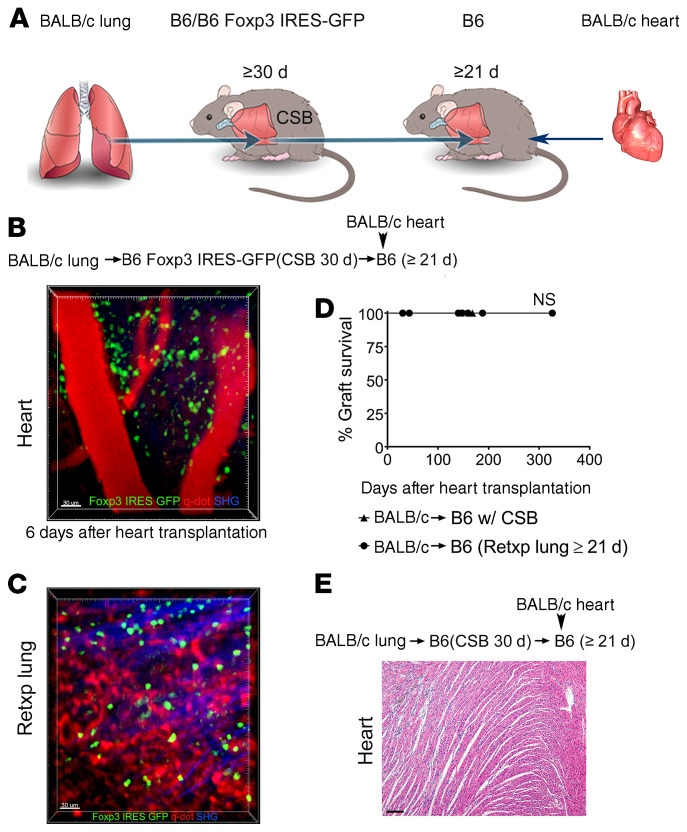
Figure 3. Peripheral tolerance is associated with exit of Foxp3+ cells from tolerant lung allograft.
(A) Schematic diagram depicting experimental transplant model. Intravital 2-photon imaging of (B) BALB/c cardiac allograft (n = 3) and (C) retransplanted (Retxp) tolerant BALB/c lung allograft (n = 3) 6 days after transplantation of BALB/c heart into B6 mouse into which a tolerant BALB/c lung was retransplanted at least 21 days prior (Foxp3+ cells: green; quantum dot–labeled [q-dot–labeled] vessels: red; second harmonic generation [SHG]: blue). The BALB/c lung had been originally transplanted into a B6 Foxp3 IRES-GFP mouse (treated with perioperative costimulatory blockade) at least 30 days before the retransplant procedure. (D) Kaplan-Meier survival curves of BALB/c hearts (▲) that were transplanted into B6 recipients that received perioperative costimulatory blockade (n = 5) or (●) that were transplanted into nonimmunosuppressed B6 mice that received tolerant BALB/c pulmonary allografts at least 21 days before cardiac transplantation (n = 7). The BALB/c lungs had been originally engrafted into B6 mice that received perioperative costimulatory blockade and then retransplanted at least 30 days later. (E) Histological appearance (H&E) of long-term-surviving BALB/c hearts after transplantation into B6 mice into which a tolerant BALB/c lung allograft was retransplanted at least 21 days prior. Scale bars: 30 μm (B and C) and 100 μm (E).

