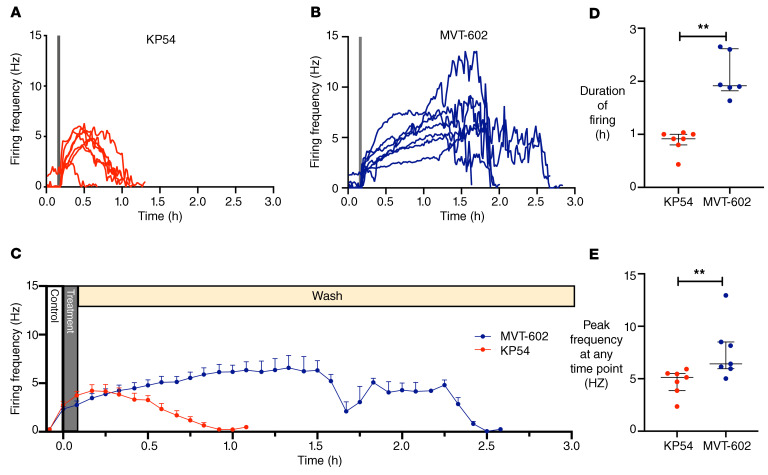
Figure 4. Targeted extracellular recordings of firing rate of GFP-identified GnRH neurons reveal that both KP54 and MVT-602 rapidly increase action potential firing rate, but the response to MVT-602 is more prolonged.
Spontaneous basal activity (control) was recorded for 10 minutes, then either 10 nM KP54 or 10 nM MVT-602 was bath-applied for 5 minutes, followed by a wash period of at least 40 minutes. (A and B) Firing frequency (Hz in 1-minute bins) of individual GnRH neurons versus time (hours) (n = 7) before, during (gray bar), and after exposure to either 10 nM KP54 (A) or 10 nM MVT-602 (B). (C) Mean (± SEM) firing frequency of GnRH neurons (n = 7 per group, 5-minute bins) over time (hours) before, during (gray bar), and after exposure to either 10 nM KP54 (red) or 10 nM MVT-602 (blue). Statistical analysis by mixed-effect model (restricted maximum likelihood) was truncated at 65 minutes because all KP54 cells had returned to baseline (P = 0.003). (D) Median (IQR) of duration of response (hours) of GnRH neurons maintaining a firing frequency greater than 1 Hz (n = 7 per group) after exposure to 10 nM KP54 (red) or 10 nM MVT-602 (blue). Groups were compared by the Mann-Whitney U test (**P = 0.0012). (E) Median (IQR) of the peak firing frequency (Hz) of GnRH neurons (n = 7 per group) at any time point after exposure to 10 nM KP54 (red) or 10 nM MVT-602 (blue). Groups were compared by the Mann-Whitney U test (**P = 0.007).

