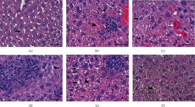
Figure 1.

Histopathological aspects of the liver. Photomicrographs of liver sections stained with hematoxylin and eosin. Bar = 50μm, 400x magnification. (a) Representative image of groups exposed to ambient air (CG, VG, QG). (b)–(e) Representative images of the lesions found in the group exposed to cigarette smoke (CSG). (f) Representative image of the group pretreated with quercetin and exposed to cigarette smoke (QCSG). (a) Preserved liver parenchyma, presence of hydropic degeneration (black arrow), and preserved sinusoid capillaries (S). (b) Hydropic degeneration (black arrow), congestion of sinusoid (black arrowhead), presence of granuloma (dotted circle), and hyperplasia (white arrowhead). (c) Hydropic degeneration (black arrow) and bleeding focus (white arrow). (d) Inflammatory cell infiltration and hydropic degeneration (black arrow). (e) Hydropic degeneration (black arrow), congestion of sinusoid (black arrowhead), and presence of granuloma (dotted circle). (f) Preserved hepatic parenchyma notes the hydropic degeneration (black arrow) and well-preserved sinusoid capillaries (S).
