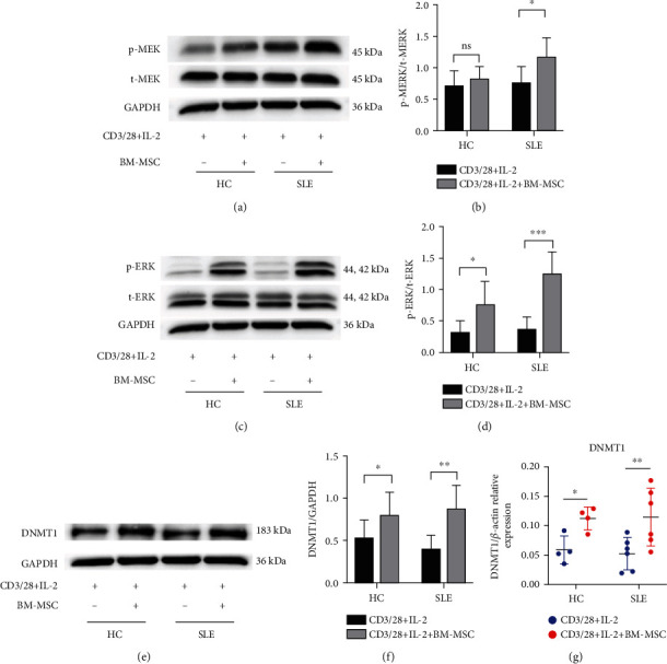
Figure 2.

MEK/ERK pathway of SLE PBMC is activated, and DNMT1 increases after the coculture with MSC. PBMC were isolated from healthy controls and lupus patients. After being stimulated with IL-2, anti-CD28, anti-CD3, both of them were divided into the single-culture group and coculture group (PBMC : BM‐MSC = 15 : 1). (a, c) The expressions of p-MEK, t-MEK, p-ERK, and t-ERK of PBMC were assessed by western blot analysis after 3 days. (b, d) The densitometry analysis of the results (a, c) is shown as the ratio of phosphorylated protein and total protein. (e–g) The protein and mRNA of DNMT1 of PBMC were determined by real-time PCR and western blot, respectively, after 4 days. The data of (a)–(d) are presented as the mean ± SD of 8 healthy volunteers and 8 patients. The data of (e)–(g) are represented as the mean ± SD of 4 healthy volunteers and 6 patients. ns: not significant. ∗P < 0.05, ∗∗P < 0.01, and ∗∗∗P < 0.001.
