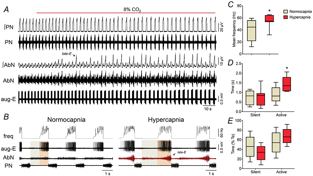
Figure 2. Changes in the discharge pattern of aug-E neurons in the BötC during the exposure to hypercapnia.

Panel A: raw and integrated (∫) recordings of phrenic (PN) and abdominal (AbN) nerve activities, and unitary recordings an aug-E neuron of the BötC of a control in situ preparation, representative from the group, illustrating the emergence of active expiration (late-E bursts in AbN, arrows) with the increase of fractional concentration of CO2 to 8% in the perfusate. Panel B: expanding recordings from panel A, illustrating the pattern of PN, AbN and aug-E neuronal activities (unit and instantaneous frequency) under normocapnia and hypercapnia. The colored boxes delineate the active and silent periods of neuronal activity. Note the increased firing frequency of aug-E neuron when AbN late-E burst emerges. Panels C-E: average values of mean firing frequency (C), and durations of active and silent periods of BötC aug-E neurons, in seconds (D) and in percentage values relative to the expiratory time (E), during normocapnia and hypercapnia (n=9). * different from corresponding values during normocapnia, P<0.05 (paired t test for data in panel C, and RM two-way ANOVA followed by Bonferroni’s post-hoc test for data in panel D and E).
