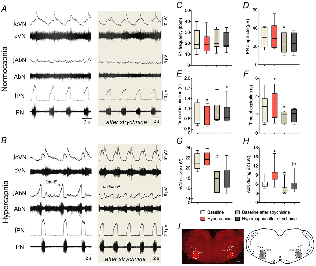
Figure 3. Strychnine microinjections in the BötC suppress the emergence of active expiration during hypercapnia.

Panels A-B: raw and integrated (∫) recordings of cervical vagus (cVN), abdominal (AbN) and phrenic (PN) nerve activities of a control in situ preparation, representative from the group, under normocapnia and hypercapnia, respectively, before and after bilateral microinjections of strychnine (10 μM) in the BötC. Panels C-H: average values of PN frequency and amplitude, times of inspiration and expiration, cVN post-I and AbN E2 activities, respectively, during baseline (normocapnia) and hypercapnic conditions, before and after strychnine microinjections in the BötC of control in situ preparations (n=7). * different from baseline, # different from baseline after strychnine, † different from hypercapnia, P<0.05 (RM one-way ANOVA followed by Bonferroni’s post-hoc test). Panel I: coronal section of the brainstem from a control in situ preparation, illustrating the sites of bilateral microinjections of strychnine in the BötC. The injection sites in each animal of the group are represented as the grey area in the schematic drawing on the right.
