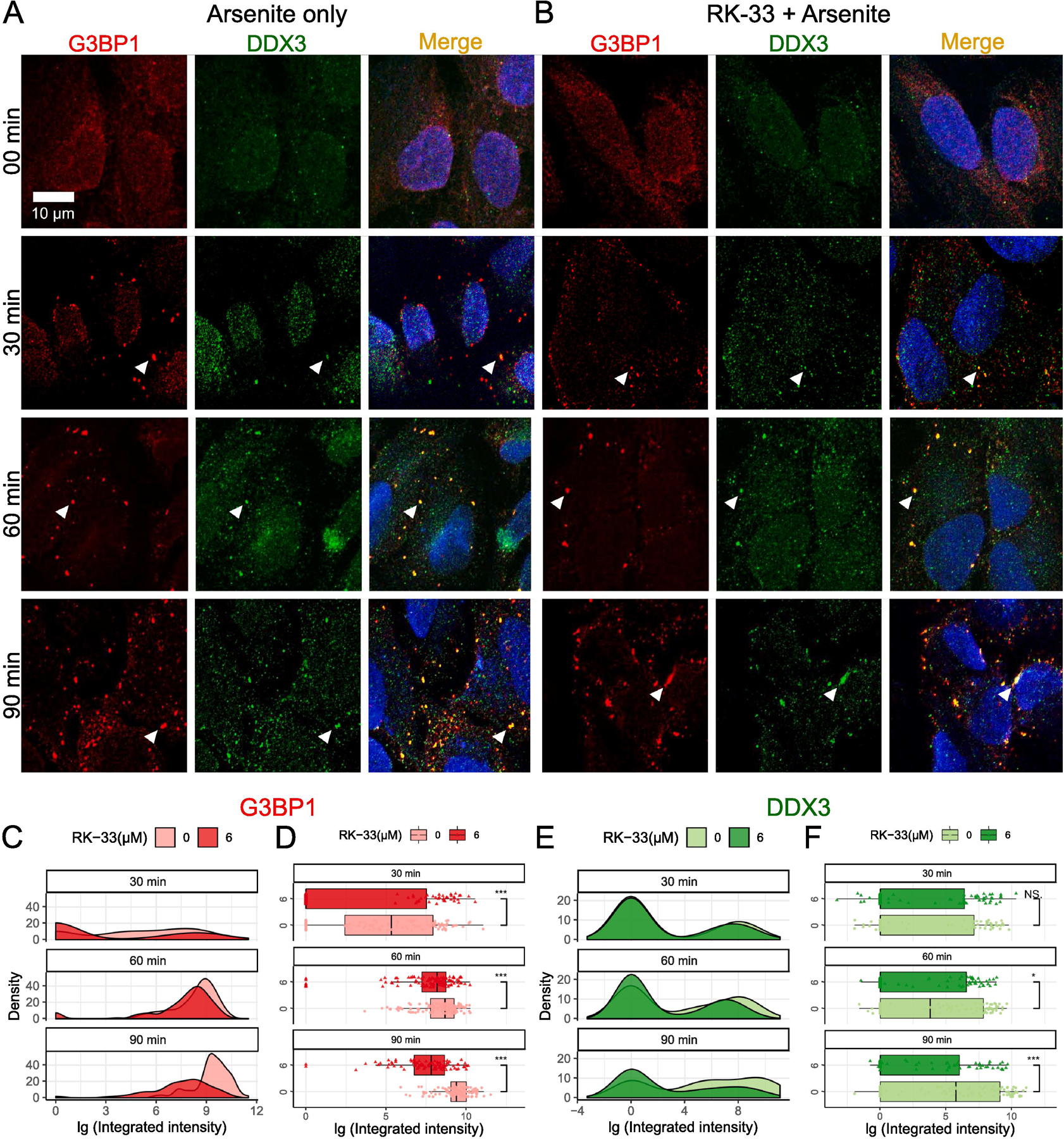
Fig. 1.

Effects of RK-33 treatment on SG assembly. (A–B) SG assembly was examined by Immunofluorescence with anti-G3BP1 (red) and anti-DDX3 (green). Scale bar, 10 µm. Cells were subjected to 00 min, 30 min, 60 min, and 90 min of 0.5 mM arsenite stress either in the (A) absence or (B) presence of 6 µM RK-33 pre-treatment for 1 h. The results of the integrated intensity are presented in log10 scale units, and graphically visualized in the form of density plots on the right and box-and-whisker plots with corresponding statistical parameters on the left. (C and D) Time course distribution of G3BP1 granules either in absence (lighter red) or presence (darker red) of RK-33 pre-treatment. (E and F) Time course distribution of DDX3 granules either in absence (lighter green) or presence (darker green) of RK-33 pre-treatment. (C and E) shows density plots and (D and F) shows box-and-whisker plots. The boxes cover 50% of data, and lines within boxes indicate median values. The Wilcoxon Signed-Rank Test was used to calculate statistical significance (* p < 0.05; ** p < 0.01; *** p < 0.001). The arrowheads represent endogenous G3BP1-dependent SGs.
