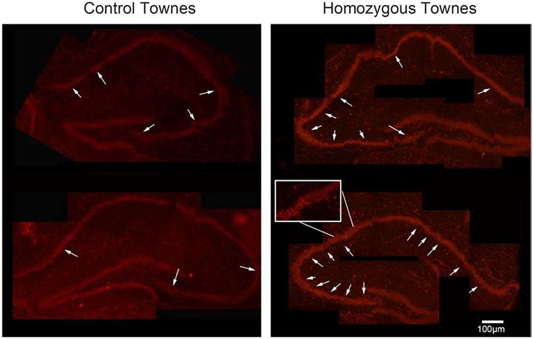
Figure 2. Townes sickle cell mice have increased superoxide levels in hippocampus.

Representative image of fluorescence indicating the presence of oxidized dihydroethidium (arrows) in hippocampi slices, a surrogate measure of reactive oxygen species production. Dihydroethidium is oxidized by superoxide to a red fluorescent material thus serving as a redox-responsive probe. Five independent raters who were unaware of samples’ genotype rated samples of hippocampi immunofluorescence (between one and four). There was high concordance in rating among the five raters (κma=0.72, SE=0.081, 95% CI: 0.57, 0.88). We found that in hippocampi from homozygous Townes, oxidized dihydroethidium staining (red color) was more intense compared to those from Townes controls (p=0.043). The intense staining was morphologically granular and reminiscent of the pyknotic neuronal morphology on H&E stained sections. Five mice were examined, three controls and two homozygotes.
