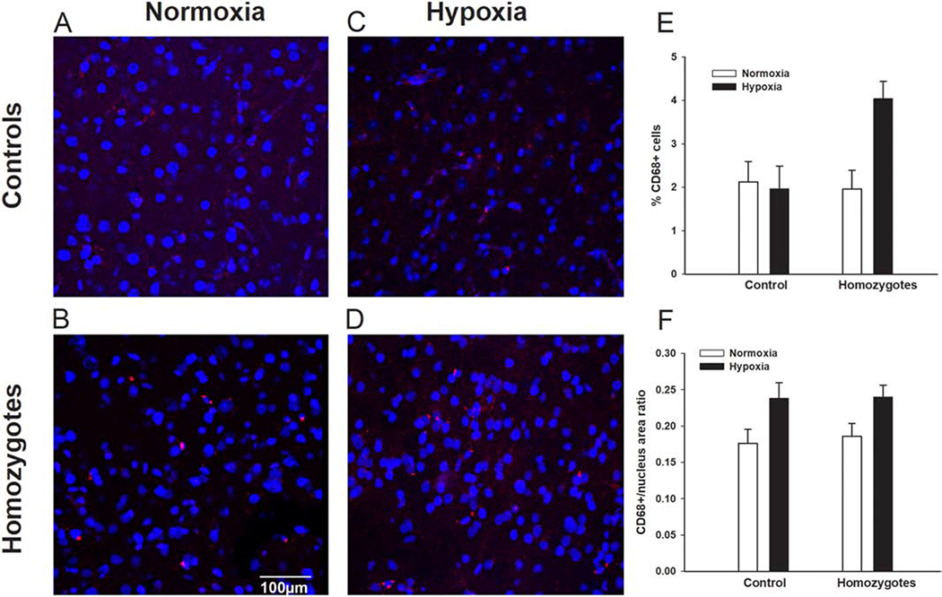
Figure 7. Hypoxia exposure is associated with microglia activation is SCD mice.

Representative images of CD68 immunofluorescence staining of cortex samples form controls and homozygous Townes after exposures to normoxia (room air) and chronic intermittent hypoxia (8% fraction of inspired oxygen)/reoxygenation for seven days. We found that during basal conditions (exposure to normoxia/room air, A and B) homozygous Townes had similar percentage of CD 68 positive cells (intense red dots, p=0.801) and similar CD68+ staining/nucleus area ratio (p=0.693) compared to controls, C, D, E, F. The effect of chronic intermittent hypoxia/reoxygenation exposures on the percentage of CD68-positive cells varied according to genotype as there was a genotype by exposure interaction (p=0.026, E). Specifically, hypoxia-exposed homozygotes, had a higher percentage of CD68 positive cells compared to normoxia-exposed animals (p=0.002). In contrast, hypoxia- and normoxia-exposed controls had similar percentage of CD68-positive cells (p=0.823). F. Overall, there was a significant effect of repeated hypoxia/reoxygenation exposures in that both controls and homozygotes had greater CD68+ staining/nucleus area ratio compared to normoxia-exposed animals (p=0.006, for effect of exposure). N=4-7 per genotype (control, homozygote) and exposure (normoxia, hypoxia).
