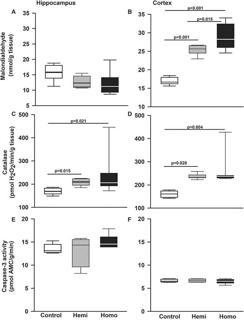
Figure 8. The BERK strain of SCD mice also has increased oxidative stress in hippocampi and cerebral cortex.

Box plots of respective variables indicate the variable’s median and first and third quartiles and the whiskers the 5th and 95th percentiles. We also examined the state of oxidative stress in a second strain of SCD mice, the BERK strain. A. In hippocampi, malondialdehyde formation, a surrogate measurement of lipid peroxidation and oxidative stress, was similar in BERKs homozygotes, hemizygotes, and controls (p=0.078 for effect of genotype). B. In cerebral cortex, homozygous BERKs had significantly higher malondialdehyde formation compared to controls (p<0.001) and hemizygotes (p=0.015). Interestingly, hemizygous Berks also had elevated levels of malondialdehyde formation compared to controls. C. In hippocampi, homozygous (p=0.021) and hemizygous (p=0.015) BERKs had significantly higher catalase levels compared to control mice. D. In cerebral cortex, compared to control mice, homozygous (p=0.004) and hemizygous (p=0.020) BERKs had significantly higher catalase levels, Figure 6D. Contrary to Townes mice, in BERKs we found no alterations in caspase-3 activity levels in hippocampus (p=0.301 for genotype effect) or cerebral cortex (p=0.391 for genotype effect), (E and F). N=5-8 mice per genotype
