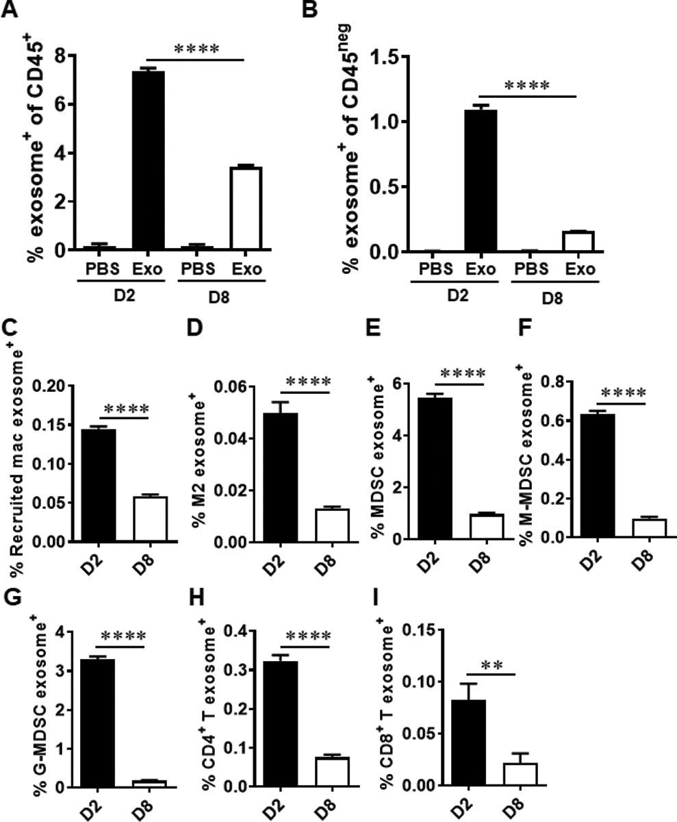
Fig. 6.

Internalization of exosomes by immune cells and tumor cells in TME. Six to eight-week-old female BALB/c mice were injected in the fourth mammary fat pad with 5 × 10⁵ 4T1.2 cells. At day 6, 7.5 × 10⁸ PKH67-labeled 4T1.2 cell-derived exosomes or PBS were injected into the tumor nodule. On day 2 (D2) and day 8 (D8) after exosome injection, tumor tissues were harvested (n = 5 mice/group). The frequencies of exosome-positive cells in CD45+ cells (a) and CD45neg cells (b) was determined by FACS analyses. The frequencies of exosome-positive recruited macrophages (c), M2 macrophages (d), MDSCs (e), M-MDSCs (f), G-MDSCs (g), CD4+ T cells (h) and CD8+ T (i) cells in CD45+ cells were determined by FACS analyses. Statistical significance was evaluated using one-way ANOVA with Tukey’s multiple comparison testing (a and b). Statistical significance was determined using unpaired t tests (c to i). ** P < 0.01, **** P < 0.001.
