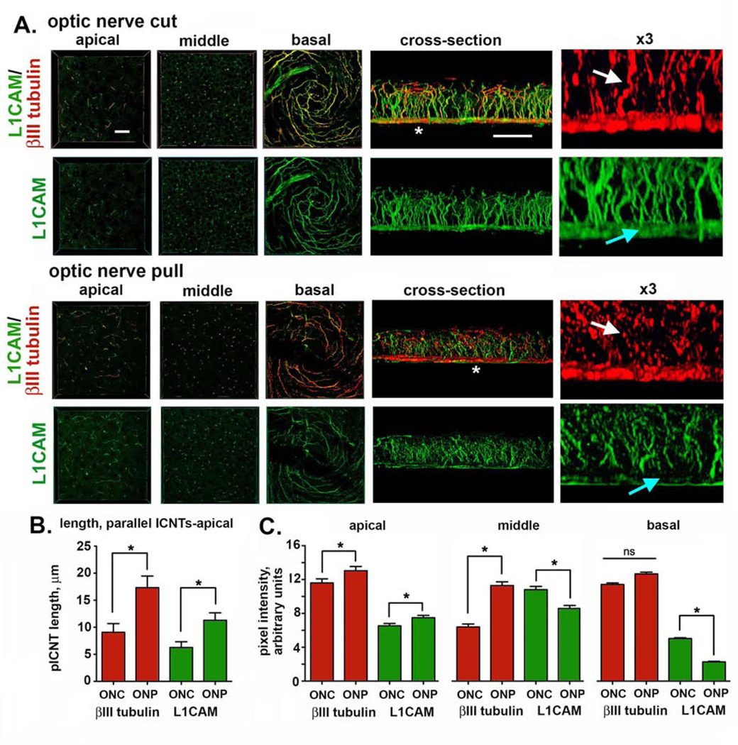
Figure 3. Enucleating eyes using the ONP method impacts ICNTs morphology and leads to shedding of L1CAM from the ICNTs.

A. High resolution confocal imaging was performed on mouse corneas enucleated using ONC or ONP after staining corneas to reveal the localization of L1CAM (green) and βIII tubulin (red). Representative en face images obtained through the apical, middle, and basal layers of the corneal epithelium are presented. 3D confocal image stacks were also obtained and rotated to allow cross-sectional view; representative cross section images are also shown. The sites indicated by the asterisks in the cross-sectional images were enlarged 3-fold and presented at the far right. White arrows show the ICNTs and highlight their beaded morphology after ONP compared to ONC. Blue arrows indicate the reduction in L1CAM at the basal aspect of the corneal epithelium where the βIII-tubulin positive ICBNs are located. Bar in the en face images = 27 μm; bar in the cross-sectional images = 25 μm. B. The average lengths of the parallel ICNTs at the apical aspect of the corneal epithelium were measured using Image J. Data show that pICNTs are longer and retain L1CAM on their surface after ONP compared to ONC. C. Cross-sectional images were used to quantify L1CAM and βIII tubulin in the apical, middle, and basal regions of the corneal epithelium. Data show that ONP significantly reduces L1CAM retention on the ICBNs and the ICNTs in the middle of the corneal epithelium but does not impact the apical -most ICNTs.
