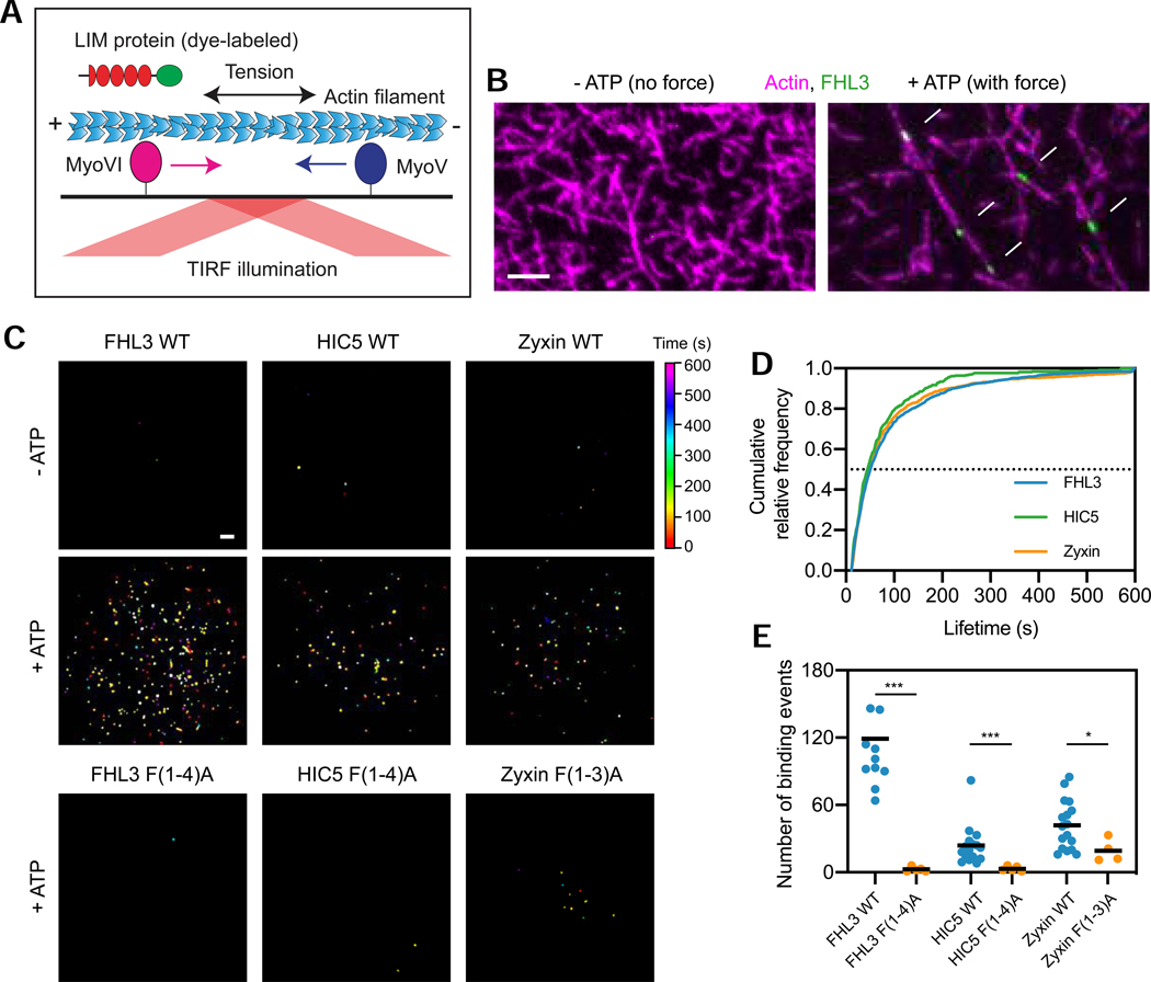
Figure 5. Strain in F-actin is necessary and sufficient for direct binding by LIM proteins.
(A) Schematic of in vitro force reconstitution TIRF assay. (B) TIRF snapshots of 10% rhodamine labelled actin filaments (magenta) and FHL3-Halo (green) in the absence (left) and presence (right) of force generation. Arrows (bottom) highlight FHL3 patches. Scale bar, 5 μm. (C) Cumulative projections of detected patches of indicated Halo-tagged wild-type constructs in the absence (top) and presence (middle) of 0.5 mM ATP, as well as mutant constructs in the presence of 0.5 mM ATP (bottom), color-coded by time. Scale bar, 10 μm. (D) Cumulative relative frequency of wild-type construct binding lifetimes. Half-lives are extracted by linear interpolation; errors represent standard deviations obtained from bootstrapping (Methods). (E) Number of patches detected in equal imaging periods across trials for wildtype and mutant constructs. Bars represent means. Dunnett’s T3 multiple comparison test after Welch’s ANOVA: * p < 0.05; *** p < 0.001. One FHL3 wild-type outlier is not shown. See also Figures S5 and S6.

