
Figure 1. Mast cell activation is associated with early neutrophil infiltration of the injured cornea.
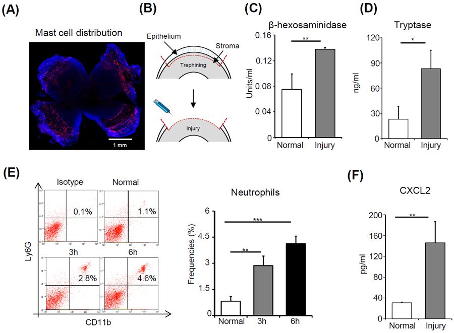
(A) Representative image showing a C57BL/6 murine cornea stained with a fluorescent conjugate of avidin (Texas Red; scale bar: 1 mm). (B) Schematic diagram depicting mouse model of corneal injury. Ocular surface tear wash was collected at 6 hours after injury and mast cell activation markers (C) β-hexosaminidase and (D) Tryptase were estimated relative to naïve mice. (E) Representative flow cytometric dot plots (left) and cumulative bar chart (right) showing the frequencies of CD11b+Ly6G+ neutrophils in the cornea at 3 and 6 hours after injury, compared to normal control animals. (F) Bar chart depicting the secretion levels of CXCL2 in the ocular surface tear wash at 6 hours following injury as estimated by ELISA, relative to normal mice. Representative data from three independent experiments are shown, and each experiment consisted of four to six animals. Data presented are mean ± SD (error bar). *p < 0.05, **p < 0.01, ***p < 0.001.