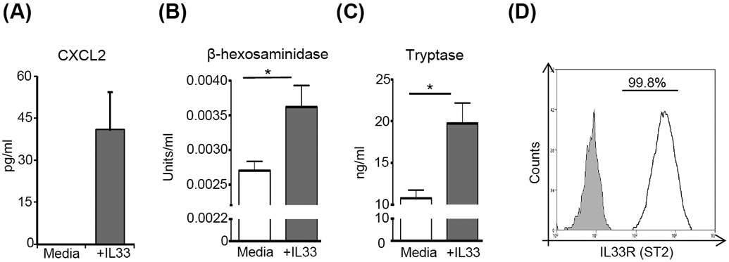
Figure 3. IL-33 activates mast cells and promotes mast cell expression of CXCL2.

(A) ELISA analysis of CXCL2 levels in the supernatants of mast cells stimulated with IL-33 compared to unstimulated cells. Supernatants from cultures of mast cells stimulated with IL-33 were analyzed for mast cell activation using (B) β-hexosaminidase (C) tryptase. Supernatant from unstimulated mast cells were used as controls. (D) Representative flow cytometry histogram showing the surface expression of IL33R (ST2; White) on mast cells as compared to isotype control (Grey). Representative data from three independent experiments are shown, and each experiment was repeated three times. Data presented are mean ± SD (error bar). *p < 0.05.
