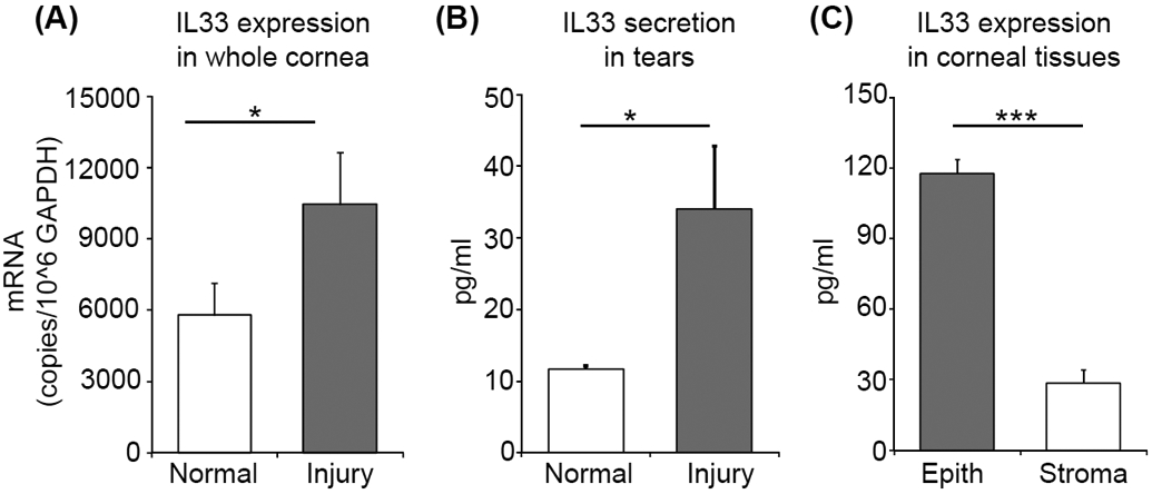
Figure 4. IL-33 released from the cornea following injury is prestored in epithelium.

(A) Bar chart depicting mRNA expression of IL-33 in lysate of whole corneas harvested at 6 hours post-injury (normalized to GAPDH) relative to naïive cornea lysate, as quantified by real-time PCR. (B) Bar chart depicting ELISA analysis of IL-33 levels in the ocular surface tear wash at 6 hours post-injury relative to naïive mice. (C) Bar chart depicting ELISA analysis of IL-33 levels in the lysates derived from separated corneal epithelium and stromal tissue lysates. Representative data from three independent experiments are shown, and each experiment consisted of four to six animals. Data presented are mean ± SD (error bar). *p < 0.05, ***p < 0.001.
