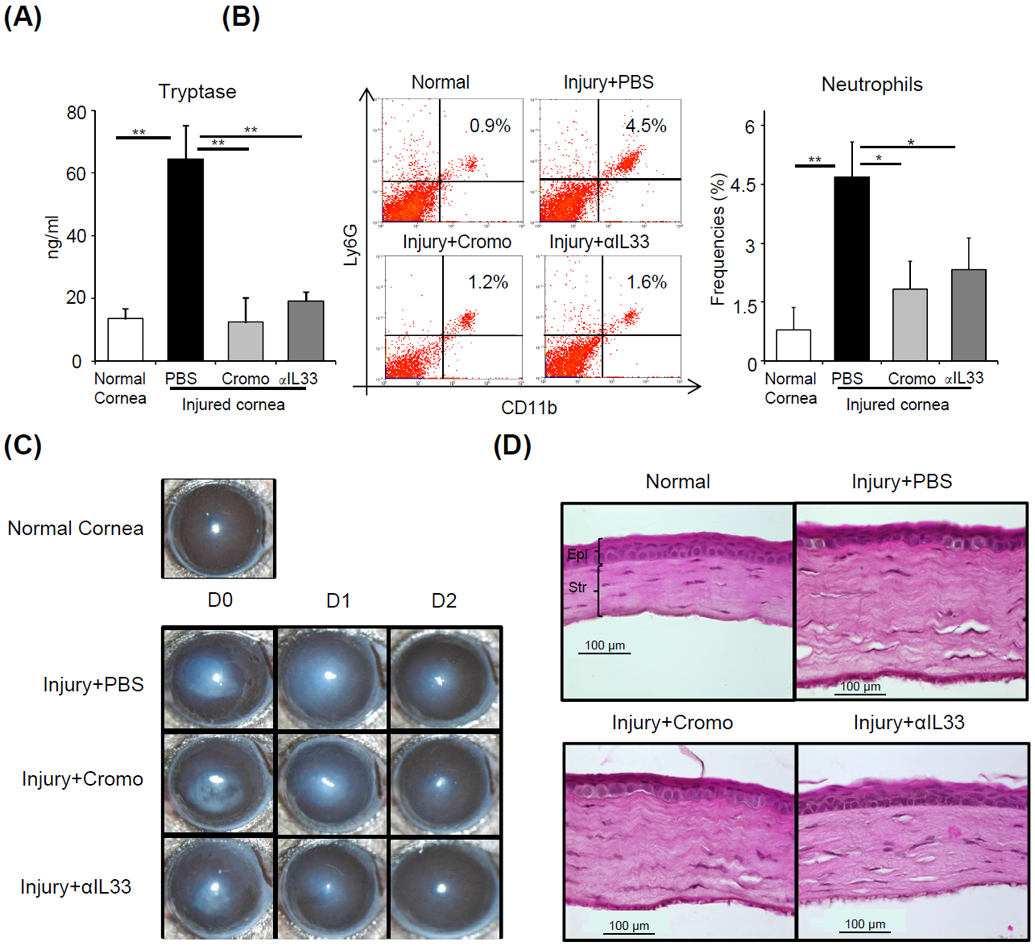
Figure 5. In vivo blockade of IL-33 suppresses mast cell activation and post-injury corneal inflammation.

C57BL/6 mice were treated topically with either cromoglycate (2%) or αIL-33 (1 mg/ml) at 1 hour prior to injury, at the time of injury as well as 1 and 3 hours after injury. (A) Bar chart depicting tryptase levels in ocular surface tear wash at 6 hours post-injury in the indicated groups relative to normal mice. (B) Representative flow cytometry dot plots (left) and cumulative bar chart (right) showing the frequencies of CD11b+Ly6G+ neutrophils in the cornea at 6 hours after injury in the indicated groups, relative to normal mice. (C) Representative bright-field microscopic images of corneas of indicated groups at Day 0, 1 and 2 post-injury. (D) Cross-sections stained with hematoxylin and eosin to visualize corneal thickness, edema and stromal lamellae (Epi: epithelium; Str: Stroma; scale bar: 100 μm). Representative data from three independent experiments are shown, and each experiment consisted of four to six animals. Data presented are mean ± SD (error bar). *p < 0.05, **p < 0.01.
