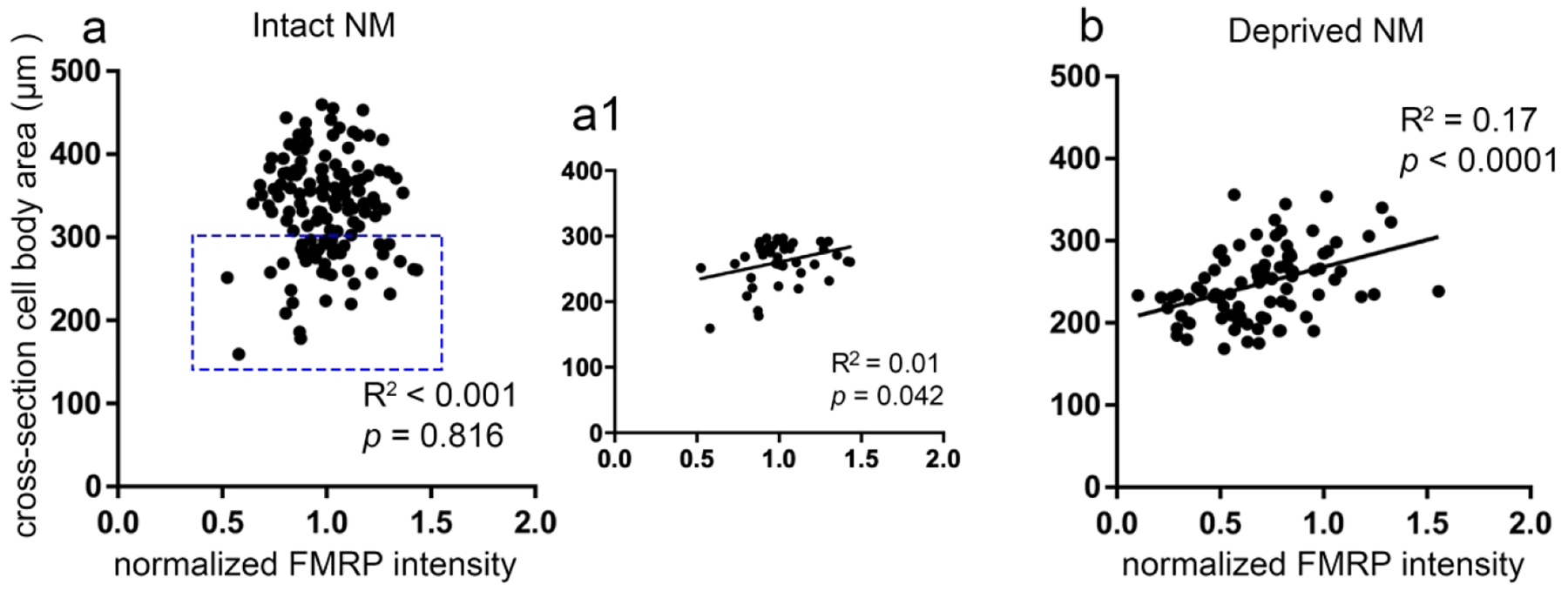
Figure 10. Correlation of FMRP intensity with cell body size at 1–2 weeks after unilateral cochlea removal.

There is no significant correlation between normal FMRP intensity and cross-section cell body area in the intact NM (a). When only including the small cells (less than 300 μm2, shown in the rectangle in a) to the correlation analysis, FMRP intensity is moderately correlated with cell size (a1). The correlation is significantly positive in the afferent-deprived NM (b). Abbreviation: NM, nucleus magnocellularis.
