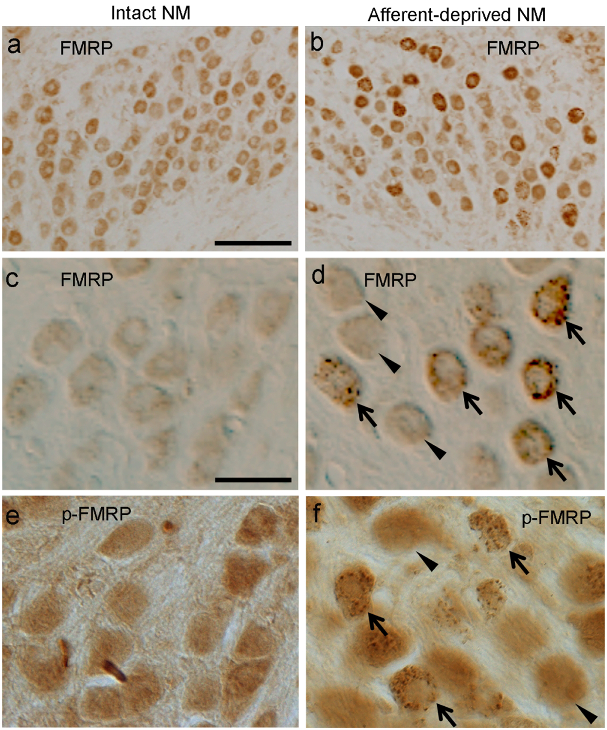
Figure 4. Peroxidase staining of FMRP and p-FMRP immunoreactivities at 6 hours following unilateral cochlea removal.

a-b: Bright-field images showing FMRP immunoreactivity in the intact (a) and afferent-deprived (b) NMs. A subset of NM neurons in b show stronger FMRP staining. c-d: Higher-magnification differential interference contrast (DIC) images of FMRP immunoreactivity in the intact (c) and afferent-deprived (d) NMs. Arrows and arrowheads point to neurons with and without FMRP puncta, respectively, in the afferent-deprived NM. e-f: DIC images of p-FMRP immunoreactivity in the intact (e) and afferent-deprived (f) NMs. Arrows and arrowheads point to neurons with and without p-FMRP puncta, respectively, in the afferent-deprived NM. Abbreviation: NM, nucleus magnocellularis. Scale bars = 100 μm in a (applies to a-b); 20 μm in c (applies to c-f).
