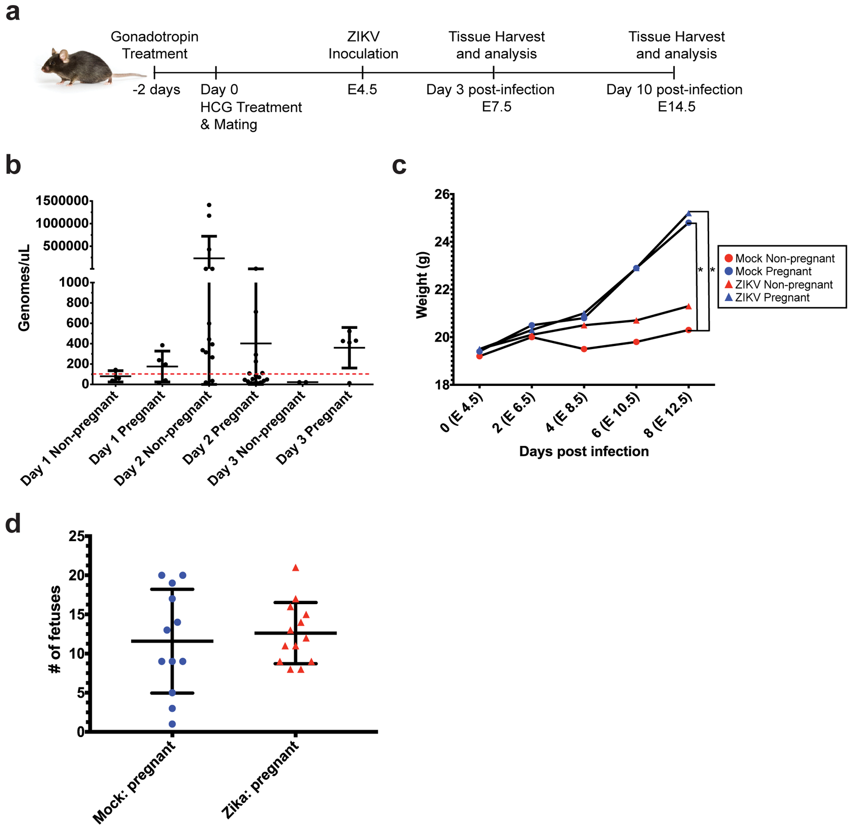
Fig 1. Intravaginal ZIKV infection in C57BL/6 mice.

a) Experimental methodology: female C57BL/6 mice were treated with gonadotropins, mated, and then infected intravaginally with ZIKV at embryonic day 4.5 (E4.5). Spleen and uterine tissues were harvested at day 3 post infection (E7.5) or day 10 post-infection (E14.5). b) Vaginal lavages were performed at 24, 48, and 72 hours post-infection, and ZIKV RNA was detected by qPCR. N=80 (4 independent experiments of 20 mice each, only samples with a Cq <36 are shown (N=42 total, N=7 Day 1, N=28 Day 2, N=7 Day 3)) c) Mice in each experimental group were weighed every other day from E4.5 (day of infection) to E12.5 (day 8 post-infection). Average weights for each group are shown. N=39 (2 independent experiments with 10 or 19 mice, N=8 mock non-pregnant, N=12 mock pregnant, N=7 ZIKV non-pregnant, N=12 ZIKV pregnant). *p<0.05 Two-way ANOVA with Tukey’s multiple comparisons. d) Whole fetuses from pregnant ZIKV-infected (red triangles) and mock-infected (blue circles) mice were dissected and counted. Each symbol (triangle or circle) represents one mouse. N=25 mice (12 mock pregnant, 13 ZIKV pregnant, taken from 2 independent experiments). Error bars represent the mean ± standard deviation in all the graphs.
