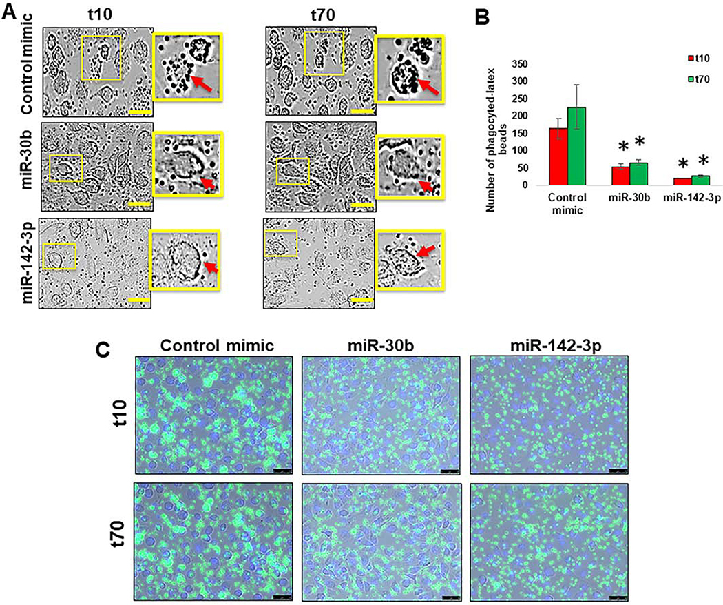
Figure 2. miR-30b and miR-142–3p transfected cells show impaired phagocytosis.
(A) Macrophages were transfected with miR-30b, miR-142–3p or control mimics and phagocytosis assay was performed after 36 h by incubating cells with FITC-conjugated IgG-coated latex beads. Representative live, contrast-phase microscopy images of phagocytosed-latex beads coated with IgG-FITC in MΦ transfected with miR-30b, miR-142–3p or control mimic were captured at 10 min and 70 min after incubation. Data are means ± SEM of three independent experiments (*p <0.05, compared with the control mimic for same time point). Scale bar- 50 μm. (B) Histograms showing number of phagocytosed beads quantified in MΦ transfected with miR-30b or miR-142–3p mimics compared with the t10 of the corresponding control mimic, and (C) a decrease of intensity and number of FITC beads were performed by live imaging in MΦ transfected with miR-30b and miR-142–3p after 36 h.

