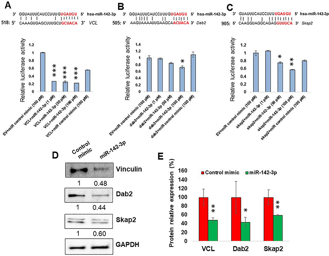
Figure 5. miR-142–3p directly regulate genes involved in cell migration, phagocytosis and cell polarity.
(A-C; Upper panel) Sequence alignment of predicted miR-142–3p binding sites in the 3′UTR of VCL, Dab2 and Skap2. Only the binding sites with mfe <−20 kcal/mol are shown. (A-C; Lower panel) HEK293 cells were co-transfected with dual luciferase reporter plasmids containing 3′UTR of VCL, Dab2 and Skap2 or control vector and miR-142–3p or control mimic. After 36 h, cell lysates were prepared to measure renilla and firefly luciferase activity. Renilla activity was normalized to firefly activity and the ratios were subsequently normalized to empty vector transfected with miR-142–3p mimic set as 1. Data are expressed as ± SEM of four independent transfections. Student’s t-test was conducted to calculate p-values. *p<0.05, **p<0.01, ***p<0.001. (D) Expression of VCL, Dab2 and Skap2 in MΦ by Western blot. Day 7 differentiated cells were transfected with miR-142–3p mimic or control mimic. Cell lysates were prepared after 36 h of transfection and VCL, Dab2 and Skap2 levels were detected by immunoblotting. The expression level of GAPDH was included as loading control. (E) The corresponding densitometric analysis by Image Lab software is also shown. Data are means ± SEM of three independent experiments (*p <0.05, **p<0.01; compared with the control mimic).

