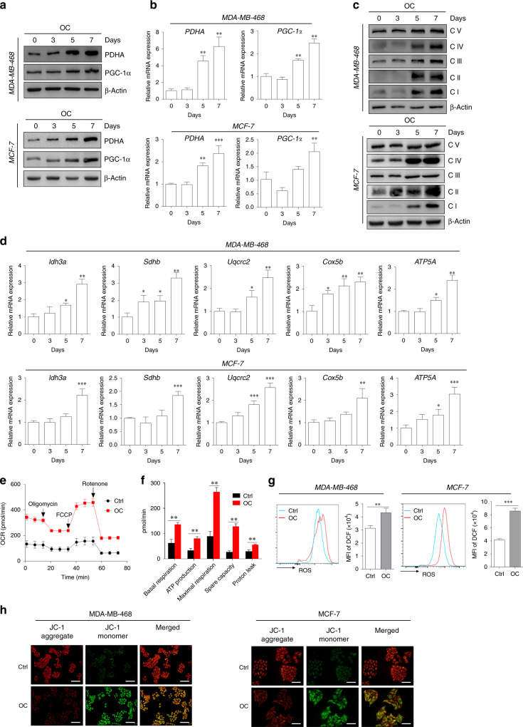
Fig. 5. Enhancement of mitochondrial metabolism in the calcification-mediated epithelial-to-mesenchymal transition (EMT) model.
The expressions of mitochondrial genes (e.g., PDHA and PGC-1α) of MDA-MB-468 and MCF-7 cells at different induction time points were evaluated by western blotting (a) and quantitative real-time (qRT)-PCR (b). The levels of OXPHOS complex subunits (c) were evaluated using western blotting at the corresponding induction time. d mRNA levels of Idh3a, Sdhb, Uqcrc2, Cox5b and ATP5A representing the OXPHOS complex were determined using qRT-PCR. β-actin was used as an internal control. e Seahorse analysis of oxygen consumption rate (OCR) in MDA-MB-468 cells at specific time points induced by OC. f OCR of basal and maximal mitochondrial respiration, ATP production, spare capacity and proton leak was analysed. g Flow-cytometry analysis of the intercellular reactive oxygen species (ROS) level by the fluorescent probe DCFH-DA in 7 days following OC treatment. h Alterations of mitochondrial membrane potential in breast cancer cells were measured by membrane-permeant JC-1 dye staining. Scale bars = 100 µm. *P < 0.05, **P < 0.01, ***P < 0.001; NS not significant, determined by unpaired Student’s t test and one-way ANOVA with Bonferroni post-test correction. Data are demonstrated as mean ± SD, and they represent three independent experiments with similar results.

