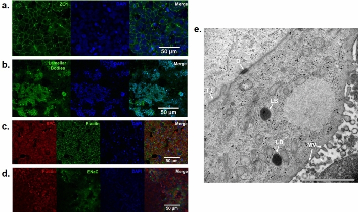
Figure 5.
Morphological characterization of the alveolar mucosa model by confocal and transmission electron microscopy (TEM). (a) The cell junction protein zona occludin 1 (ZO1) (b) lamellar bodies, (c) surfactant protein C (SPC) and (d) epithelial sodium channel (ENaC). Nucleus is stained in blue. Bar scale: 50 µm; (e) Representative TEM image of the alveolar type II cells in air–liquid interface (2 weeks) showing microvilli (MV), lipid bodies (LB), desmosome (D), and tight junction (TJ) Bar scale: 2 µm. The microscopic images are representative of alveolar mucosa model developed at air–liquid interface (2 weeks) from NCl-H441 cells.

