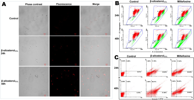
Figure 6.
(A) Superfluous lipid body detection in L. donovani promastigotes. Promastigotes (1 × 10⁷/ml) treated with β-sitosterolCCL for 24 h and 48 h; images were obtained by LSM510-META confocal microscopy after Nile Red staining. Phase contrast, fluorescence, and phase contrast-fluorescence merged images are representative of three independent experiments. (B) Flow cytometric determination of mitochondrial membrane depolarization. Promastigotes (1 × 10⁷/ml) treated with β-sitosterolCCL (IC50 dose) for 24 h and 48 h followed by JC-1 staining. The fluorescence intensity of JC-1 was measured, and histograms (snapshot) of β-sitosterolCCL-treated parasites were compared with control and miltefosine (10 µM)-treated cells for 24 h and 48 h. Red (R-1 region) and green (R-2 region) depict high and low mitochondrial membrane potential, respectively. The snapshots are representative of one of three independent experiments. Data were acquired in a BD FACSCalibur flow cytometer and analysed in Flowing software (https://www.flowingsoftware.com), version 2.5.1, Finland. (C) Determination of externalization of phosphatidylserine by flow cytometry. Promastigotes (1 × 10⁷/ml) were treated with β-sitosterolCCL followed by annexin V-FITC and PI staining. Respective dot plots of β-sitosterolCCL-treated parasites are represented along with control and miltefosine (10 µM)-treated cells for 24 h and 48 h. Data were acquired in a BD FACSCalibur flow cytometer and analysed in Flowing software (https://www.flowingsoftware.com), version 2.5.1, Finland.

