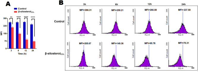
Figure 7.
Measurement of intracellular non-protein thiols in promastigotes. Promastigotes (1 × 10⁷/ml) were treated with β-sitosterolCCL, and fluorescence intensity was measured after CMFDA staining at various time points, i.e., 3 h, 6 h, 12 h, and 24 h. (A) The bar graph depicts the MFI values of three independent experiments, and statistical significance was determined with respect to the control by using a t-test, where **p < 0.01 and ****p < 0.0001 were considered statistically significant. (B) Histograms depicting the reduction of MFI in β-sitosterolCCL-treated parasites compared to the control at every time point. Data were acquired in a BD FACSCalibur flow cytometer and analysed in Flowing software (https://www.flowingsoftware.com), version 2.5.1, Finland.

