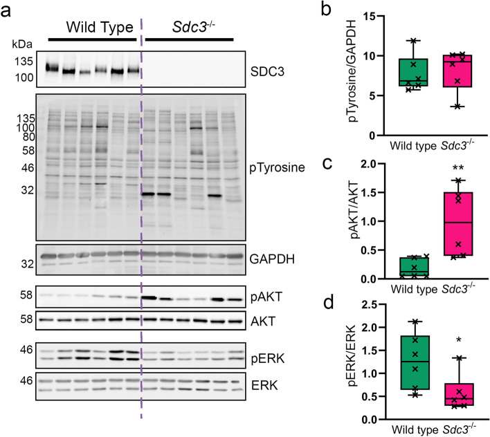
Figure 3.
Loss of SCD3 in MSCs causes increased ATK phosphorylation and a reduction in ERK phosphorylation. MSCs were isolated from wild type (n = 6) and Sdc3−/− (n = 6) mice, cultured in full growth medium, and lysed ready for western blotting. (a) Representative images of blots used for quantification. SDC3 knockout was confirmed for Sdc3−/− MSCs in each sample. (b) Total phosphorylated tyrosine normalised to GAPDH. (c) Phosphorylated AKTSer473 was normalised to total AKT. (d) Phosphorylated ERKThr202/Tyr204 was normalised to total ERK. Quantification was performed using ImageJ. Data displayed as mean ± standard deviation; *p < 0.05; **p < 0.01. Full-length blots are presented in Supplementary Figure 1.

