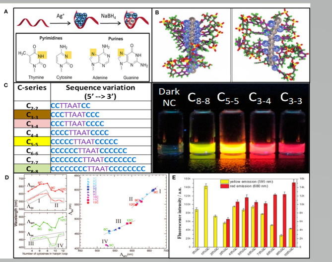
Figure 1.
(A) The red band, blue balls and gray balls representing the ssDNA template, Ag+, and Ag0, respectively. Pyrimidine N3 and purine N7 interacting with Ag+ are shown in yellow (New et al., 2016). (B) The rod-like model of the AgNCs with neutral Ag atoms represented by gray balls and Ag+ cations represented by blue balls in the poly(C) DNA. Left: in repeat tetramer units. Right: in repeat trimer units (Schultz et al., 2013). (C) Varying the length of the C base leads to AgNCs varying in color (Obliosca et al., 2014). (D) The number of C in the hairpin ring determining the wavelength (left) and the excitation vs. the emission wavelengths (right) (O'Neill et al., 2009). (E) The fluorescence intensities of the hairpin-AgNCs synthesized by different percentages of GC content in the stem sequences (Guo et al., 2020).

