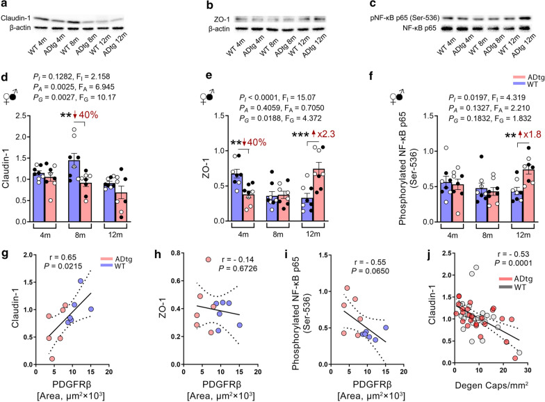
Fig. 4.
Altered expression of endothelial cell junction molecules and increased NF-κB phosphorylation in the retinas of APPSWE/PS1ΔE9 (ADtg) mice. a–c Representative Tris–glycine gel images showing western blot protein bands of claudin-1, zonula occludin-1 (ZO-1), phosphorylated NF-κB p65 subunit (pNF-κB p65) at serine-536, and total NF-κB p65 subunit as well as β-actin controls from retinal lysates of ADtg mice and age- and sex-matched wild type (WT) littermates (n = 8 for each age and genotype). d–f Densitometric analyses of western blot protein bands of d claudin-1, e ZO-1 and f pNF-κB p65, normalized by d–e β-actin control or f total NF-κB p65 in the same mice cohort. g–i Pearson’s coefficient (r) correlation between retinal PDGFRβ immunoreactivity and the densitometric analysis of western blot protein bands of g claudin-1, h ZO-1, or i pNF-κB p65 in a subset of this mice cohort (n = 12). j Pearson’s coefficient (r) correlation between degenerated retinal capillary count and the densitometric analysis of western blot protein bands of claudin-1. Data from individual mice (circles) as well as group means ± SEMs are shown. Black-filled circles represent males and clear circles represent females. Fold or percentage changes are shown in red. **p < 0.01, ***p < 0.001, by two-way ANOVA with Tukey’s post hoc multiple comparison test. P and F values of two-way ANOVA refer to comparisons of age groups (PA, FA), ADtg versus WT genotype groups (PG, FG), interactions (PI, FI)

