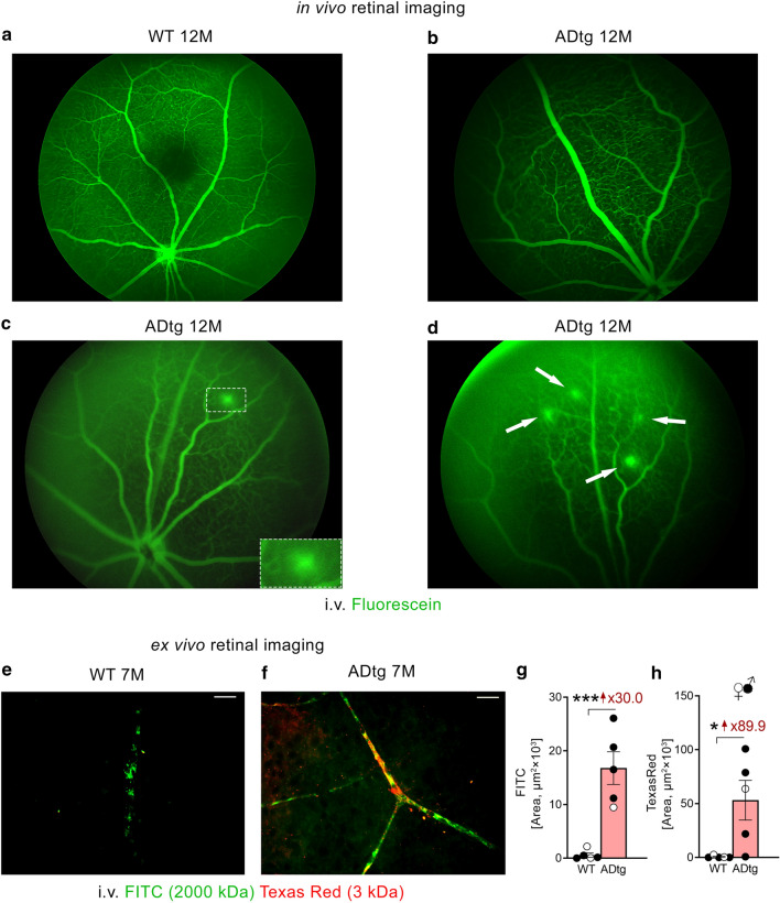
Fig. 5.
Retinal microvascular leakage in APPSWE/PS1ΔE9 (ADtg) mice. a–d Representative images of noninvasive retinal microvascular imaging after intraperitoneal fluorescein injection in 12-month-old a wild type (WT) and b–d ADtg mice. Note: images in b showing intact retinal microvasculature and in c–d showing leaked retinal microvasculature. e–f Representative images of retinal flat-mount obtained from 7-month-old WT and ADtg mice (cohort average age is 6 month) that received intravenous tail injections of FITC-dextran and Texas Red-dextran. g–h Quantitative analysis of the FITC or Texas Red-stained area in each microscopic field of retinal flat-mounts from WT (n = 5) or ADtg (n = 5) mice. Black-filled circles represent males and clear circles represent females. *p < 0.05, ***p < 0.001, by an unpaired 2-tailed Student t-test. Fold changes are shown in red

