Figure 7.
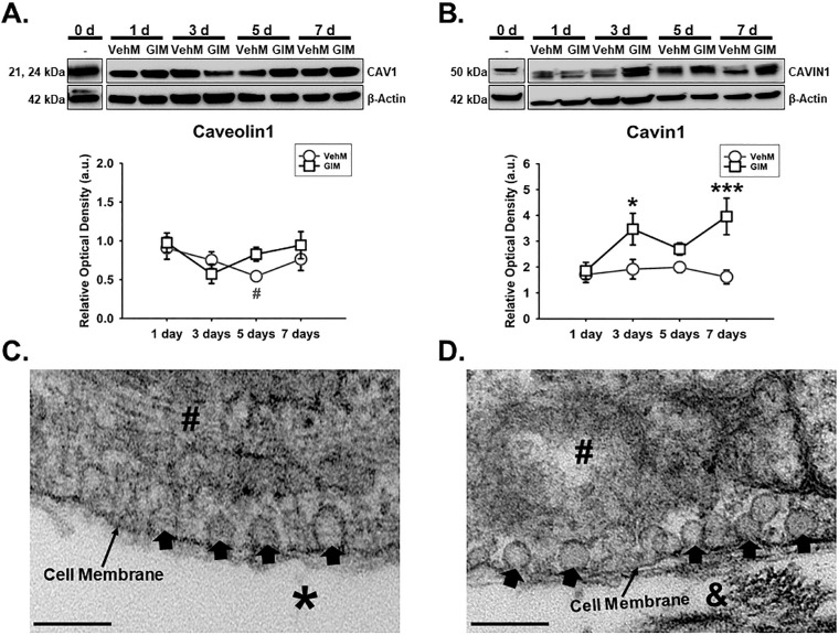
VehMs and GIMs form caveolae with GIMs temporally overexpressing Cavin1 in hTM cells. Primary hTM cells were treated with vehicle control (Veh) or 100 nM dexamethasone (DEX) in complete growth media for 4 weeks to obtain VehMs and GIMs, respectively, following successful decellularization. Fresh, earlier passage, hTM cells from the same donor used to obtain these matrices were cultured on VehMs and GIMs in 1% fetal bovine serum (FBS) growth media for 1, 3, 5, and 7 day(s). Protein was extracted for Western blot analysis, and transmission electron microscopy was performed to visualize caveolae. Data from VehM and GIM were respectively normalized to baseline protein levels (time point 0 day). β-Actin was used as an internal control. Respective representative blot (top) and densitometric analysis (bottom) of (A) Caveolin1, and (B) Cavin1. Representative electron micrograph demonstrating cell membrane-bound caveolae in cells cultured on (C) VehMs and (D) GIMs for 7 days. Columns and error bars; means and standard error of mean (SEM). Two-way ANOVA with the Holm Sidak pairwise comparisons post hoc test was used for statistical analysis (n = 5 biological replicates). *P < 0.05, ***P < 0.001 for GIM versus VehM, given significant treatment and time interaction. #P < 0.05 VehM versus baseline protein. hTM, human trabecular meshwork; VehMs, vehicle control matrices; GIMs, glucocorticoid-induced matrices. Arrow heads, Caveolae; #, Cytoplasm; *, VehM; &, GIM. Scale bar, 200 nm.

