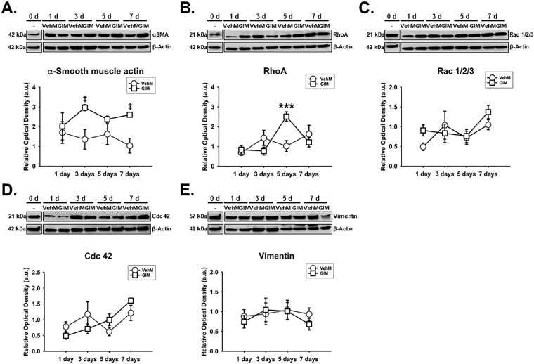
Figure 8.
GIM temporally and differentially upregulated specific actin cytoskeletal-related proteins hTM cells. Primary hTM cells were cultured in the absence or presence of 100 nM dexamethasone (DEX) in complete growth media for 4 weeks and decellularized to obtain VehMs and GIMs respectively. New hTM cells from the same donor used to derive these matrices were cultured on VehMs and GIMs in 1% fetal bovine serum (FBS) growth media for 1, 3, 5, and 7 day(s). Protein was extracted for Western blot analysis. VehM and GIM were, respectively, normalized to baseline protein levels (time point 0 day). β-Actin was used as an internal control. Respective representative blot (top) and densitometric analysis (bottom) of (A) α-smooth muscle actin, (B) RhoA, (C) Rac 1/2/3, (D) Cdc 42, (E) vimentin. Columns and error bars; means and standard error of mean (SEM). Two-way ANOVA with the Holm Sidak pairwise comparisons post hoc test was used for statistical analysis (n = 5 biological replicates). ***P < 0.001 for GIM versus VehM, given significant treatment and time interaction. ‡P < 0.05 for GIM versus VehM, given significant main effect of treatment). hTM, human trabecular meshwork; VehM, vehicle control matrix; GIM, glucocorticoid-induced matrix.

