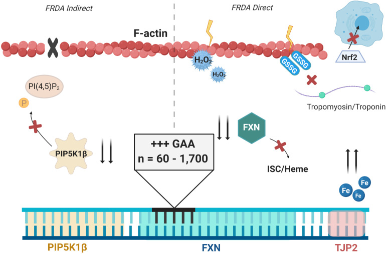
FIGURE 3.
A visual representation of ideas highlighted in this review concerning altered actin physiology in FRDA. The root of these alterations is the increase in GAA expansions in the first intron of FXN, shown in blue. Decreased transcriptional processing of PIP5K1β shown in yellow prevents phosphorylation of PI(4,5)P2, preventing normal actin polymerization (left, indirect effect of FRDA). Decreased production of FXN prevents normal ISC and heme biogenesis, permitting free cellular iron. Free radical production and oxidative stress follows, causing direct oxidation of actin filaments and glutathione addition, which prevents polymerization and binding to tropomyosin for stabilization (right, direct effect of FRDA). Nrf2 is shown to have inhibited nuclear translocation. TJP2 also flanks FXN as shown in pink, but is not known to be altered in FRDA at this time.

