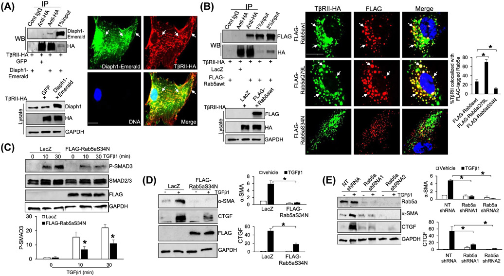
FIGURE 6.
TβRII interacts with Diaph1 and Rab5a and inactivation of Rab5a suppresses TGFβ1-stimulated HSCs activation. A, Left, HSCs expressing TβRII-HA and Diaph1-Emerald were collected for IP and HSCs expressing TβRII-HA and GFP were used as a control. Anti-HA was used to pull down TβRII-HA and coprecipitated Diaph1-Emerald was detected by WB using anti-Diaph1. TβRII-HA interacted with Diaph1-Emerald in HSCs. Right, double IF revealed that TβRII-HA and Diaph1-Emerald colocalized at the plasma membrane and endocytic vesicles (arrows). Bar, 20 μm. B, Left, HSCs expressing TβRII-HA and FLAG-Rab5awt were collected for IP and HSCs expressing TβRII-HA and LacZ were used as a control. Anti-HA was used to pull down TβRII-HA and coprecipitated FLAG-Rab5awt was detected by WB using anti-FLAG. TβRII-HA interacted with FLAG-Rab5awt in HSCs. Right, HSCs coexpressing TβRII-HA and FLAG-tagged wild-type Rab5a or a mutant were subjected to double IF. FLAG-Rab5aQ79L increased whereas FLAG-Rab5aS34N reduced TβRII-HA/Rab5a colocalization, compared to FLAG-Rab5awt (arrows). *P < .05 by ANOVA, n = 6 cells per group. Bar, 20 μm. C and D, HSCs expressing LacZ (control) or FLAG-Rab5aS34N were stimulated with TGFβ1 for indicated times and collected for WB for p-SMAD3, α-SMA, and CTGF. Expression of Rab5aS34N suppressed TGFβ1-mediated phosphorylation of SMAD3 and HSC activation. *P < .05 by ANOVA, n = 3. E, HSCs expressing NT or Rab5a shRNA were stimulated with TGFβ1 for 24 hours and collected for WB for α-SMA and CTGF. Knockdown of Rab5a inhibited TGFβ1-mediated HSC activation. *P < .05 by ANOVA, n = 3 repeats

