
FIGURE 2.
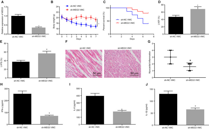
Myocarditis is relieved by decreasing MEG3 in viral myocarditis (VMC) mice. VMC mice were injected with sh‐NC or sh‐MEG3. A, MEG3 expression in myocardial tissues of VMC mice determined by RT‐qPCR. B, Change of mouse body weight. C, Survival rate of mice. D, LVEF determined by transthoracic echocardiography. E, LVFS examined by transthoracic echocardiography. F, H&E staining of myocardial tissues (×200). G, Evaluation of myocarditis according to H&E staining. H/I/J, IFN‐γ, IL‐6 and IL‐1β contents in cardiac tissues monitored by ELISA. The measurement data were described as mean ± SD. Statistical comparisons between two groups were analysed by unpaired t test, and repeated measurement ANOVA was used for comparing mice weight at different time point, followed by Bonferroni post hoc test. *P < 0.05 vs VMC mice injected with sh‐NC. N = 5 or 10. The experiment was performed at least three times